Friday, December 17, 2021

Fat embolism

Definition: 

Fat emboli:

- a complication of trauma/ surgery that involve intrumentation of femoral intramedullary canal

- it is a response/ manisfestation of fat globules that may enter the blood stream

Fat embolism:

- a process by which fat emboli passes the bloodstream and lodges within the blood vessel

Fat embolism syndrome:

- serious manisfestation of fat embolism that causes multi system dysfunction


Causes: 

1. mechanical theory

- fat droplets from bone marrow enters the vessel

- increase of intramedullary pressure and cause fat/marrow to enter the bloodstream. which later could lodge in the lungs as emboli

- causes inflammation and local ischemia

2. metabolic theory

- stress from trauma that causes change to the chylomicron that causes formation of fat emboli

or

1. trauma related

- fracture at long bones: especially femur fractures

2. non trauma related

- liver disease, shock, bone tumor lysis


Symptoms:

- asymptomatic within the first 12-48 hours

Gurd's criteria: 2major 1 minor or 1major 4 minor

  • Major: 

- hypoxemia, petechial rashes, neurological symptoms, pulmonary edema

  • Minor: 

-tachycardia, fever, retinal changes/ renal changes/ fat macroglobinemia, jaundice, 

- drop in Hb, increase ESR, thrombocytopenia


Investigation: 

- FBC

- ABG

- RP/LFT

- CXR : ground glass appearance / snow storm appearance


Management:

- stabilise the patient

  • Airway : no obstruction
  • Breathing: oxygen support
  • Circulation: two large branulla with fluid support 
    • hemodynamically: Hb? any blood loss
    • hydration: 3L/d

1. monitor vital signs : BP, PR, SPO2, temperature

2. inform 

- MO incharge, anaest (ventilator support)

- keep in view the need of doing CT brain to exclude other causes

3. rigid fixation of fracture within 24 hours

4. diagnosis of exclusion

5. DVT prophylaxis 

6. stress ulcer prophylaxis


Reference: 

1. https://www.orthobullets.com/basic-science/9055/fat-embolism-syndrome

Compartment syndrome

Definition:

a rise in pressure within a closed osteofascial compartment, resulting in impaired local circulation compartment. 


Causes:

high risk injury:

- fracture that causes inflammation to the muscle where it has no place to expand therefore compressing on the vascular structure and nerves

- operation

- infection

- trauma: crush injury

- burn injury

- tight plaster cast


clinical features

5P s

  • PAIN: pt will complain excrutiating pain, just like they wanted to have their legs cut off
  • Pallor
  • Parasthesia
  • Pulselessness
  • Paralysis

Passive stretch test: positive 

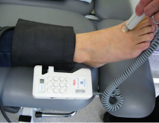
- can be seen in the video below 2:50 onwards


transducer : to measure the pressure we have at the compartment


Management : 

1. triage

2. if there is a cast or bandage, remove it immediately

**no compression

3. elevate the limb but not above heart treatment

4. emergency fasciotomy

2. Emergency Fasciotomy 

NF and GG


Necrotising Fasciitis vs Gangrene


** REMEMBER TO ALWAYS DO RESUSCITATION FIRST PRIOR OTHER MANAGEMENT (ABX, ANALGESICS, DM CONTROL)

Indication for amputation: 3D's

🐣 Dying / diseased bone - PVD, infection, gangrene, trauma
🐣 Dangerous - lethal sepsis, malignant
🐣 Damn nuisance - gas gangrene, crush syndrome, severe malformation


Sample Xray


 Gas shadows seen extending from the foot up to upper calf 


Doppler 


Usually medical officer will ask for a doppler done and also ABSI. 
So try to get your doppler right. 
There are three spots that we must check : the Dorsalis Pedis Artery (DPA), Posterior Tibial Artery (PTA) and Brachial 

                   Normal ABSI: 1.0-1.3                                                    Normal wave: Triphasic
          ABSI 0.5-0.9: moderate to severe
ABSI >1.3: common in DM (may have severe calcifications)


Antibiotics:

For NF: commonly polymicrobial bacteria can be seen. 
  • - IV Tazocin 4.5g QID (consist of piperacillin and tazobactem)
  • - IV clindamycin 600-900mg  TDS 
      • inhibitor of protein synthesis and protein  m synthesis 
      • effective for gram +ve
  • or Cefotaxime + metronidazole
 OTHERS:
  • - unasyn (beta lactam, inhibiting bacterial cell wall synthesis)
  • - ciprofloxacin : covers gram negative
Content Placeholder 4

Reference


Short terms;
- WD: wound debridement
- h/o : history of
- DM : Diabetes Mellitus
- DFU : Diabetes Foot Ulcer
- ABSI : Ankle Brachial Index

Monday, November 15, 2021

HPB Q&A for HO

HPB = Hepatobiliary

Anatomy of HPB 


Common classifications used in HPB

1. Ranson criteria


2. Mirrizi classification


biliary colic -> acute cholecystitis -> acute cholangitis, pancreatitis, gb empyema and gangrenous gb

presentation of each spectrum

Causes of obstructive jaundice


painless vs painful jaundice

  • Painful obstructive jaundice is usually related to gallstones, while painless obstructive jaundice tends to be related to tumours.


Q1: If patient came with features of ascending cholangitis, what Investigation you would like to order

  • FBC - to monitor TWC, PLT. 
    • low platelet: sepsis induces/ severe sepsis that causes coagulopathy
  • BUSE - urea, creat monitoring
    • hyperbilirubinemia can cause hepatorenal syndrome and renal impairment
    • severe sepsis that causes organ failure : kidney failure
  • LFT: TB, kiver enzymes and albumin level
    • especially ALP and direct bilirubin that can help determin if there are any obstructive jaundice
  • ABG: any acidosis or resp failure
  • CXR first, as we can use it to rule out perforated viscus
    • unless obvious jaundice, so less likely perforated PUD

Q2: why we need ultrasound HBS for ascending cholangitis?

    • to rv CBD(common bile duct) size, and any dilatation
    • any stone present- if there's obstruction , might need urgent intervention to relieve obstruction
    • sometimes stone cant be seen as obscured by bowel gas. 
      • stone is usually in distal CBD - the narrowest part
      • and it is at the back of duodemun
      • usg wave cannot go through gas
    • To rule out liver abscess
      • as the presentation of liver abscess is the same: RHC pain, fever and jaundice

Q3: normal size of CBD?

  • 3-5mm
  •  >8mm considered dilated (usually we use >10mm or 1cm as dilated)

Q4: why do we need to rule out liver abscess urgently?

  • if huge liver abscess need to drain urgently. 
  • gold standard tx: percutaneous drainage and antibiotic
  • otherwise it will rupture and cause peritonitis --> severe sepsis --> mortality

Q5. if found out patient have ascending cholangitis secondary to choledocholithiasis, how would you manage?

  • resuscitate and stabilize the patient first with ABC
  • Airway
  • Breathing
  • Circulation
  • insert branula + blood for Ix and blood culture
  • fluid resus
  • antibiotics
  • analgesia

Q5a. what are the choice of antibiotics use for the pt above?

  • HBS common organism are
  • function of liver: it uses portal circulation, 
    • it collects blood from gut  - all the guy -> return back to liver
  • therefore in the GI tract, mainly organism gram negative and anaerobes
  • also called BROAD SPECTRUM of negative organisms
    • so we need at least 3rd generation cephalosporin 
    • eg: cefobid / cefoperazone
  • to cover anaerobes: metronidazole

Q6. after analgesics what can we do for patient with ascending cholangitis and in pain?

  • if Stable, we can try ERCP to remove the stone . if unable to do so we will put stent to drain the obstruction
    • ERCP needs sedation, and prone position. therefore difficult to maintain airway
  • If patient is unstable , the option is PTBD

Q7. when to do cholecystectomy?

  • once sepsis is resolved
  • stone not cleared through ERCP 

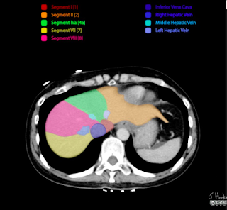
Q8. How do you tell which liver segment was it from a CT scan?

Anatomy of liver :
note that 2 is beside 4a, 3 below 2 and 4b is below 4a
upper segment at right side is segment 7,8
and lower right is segment 6 and 7


 First you can check the segments from identifying the hepatic veins. As shown in diagram above, left hepatic vein separates segment 2 and 4a, then the middle hepatic vein for segment 4a and 8, right hepatic vein for segment 8 and 7


Then as you scroll down the ct scan, you will see the portal vein (left branch). the portal vein separates the liver to the left(segment 2 and 4a) and right side (segment 7,8) . below the portal vein emerges segment5


When you can see the right branch of portal vein, it also indicates that on Left (segment 4b, and segment 3), on its right is segment 5 and segment 6


reference, more on website: 


Saturday, November 6, 2021

Electrolyte Imbalance QnA for HO

 House Officers must know in ward 

- Electrolyte imbalance


Q1: 50year old, 70 kg, male with persistent diarrhea and vomiting for 3days due to food poisoning. He is dehydrated. Vitals are stable, looks lethargic. Na: 128mmol/L

a. How to correct his hyponatremia

    - calculate first
  • Na deficit (mmol) : (desired Na level - serum Na) x bw x fraction
  • 0.6 x kg x (desired Na - serum Na)
    • desired Na :  135-145
    • Na deficit is 0.6 because extracellular comprises 60%. 
    • If its potassium (intracellular) therefore 0.4 cause it is 40%
  • 0.6 x 70 x (135-128) = 294mmol/L
    - to add with daily requirement
  • infusion = deficit + requirement
    • requirement: 1-2mmol/kg/d (we usually use the lower range when calculating)
  • infusion: 294 + 70 = 364 mmol/L
- then how do we deliver 364mmol/L to pt?
  • 1 pint NS = 77mmol/ L
  • 1L NS = 154mmol/L
  • 364 / 77 = 4.7 pints, 
  • therefore 5 pints of normal saline over 24 hours is needed.

b. the next day his repeated Na is normal: 136, 

how do you plan for fluid regime ? for maintenance?

  • normally a person need 30-40ml/kg/d (fluid requirement)
    • general normal weight for man: 70kg,  normal weight for woman : 60kg
  • if we take the middle value fluid requirement (35ml/kg/d), 
    • the man will need 2450ml (around 5 pint as well)
    • women is 2100ml, around 4pint. 
thats why usually we see usually 
  • 5 pint (2/3 pint NS + 2/3 pint D5%) given to man, 
  • 4 pint(2 pint NS + 2 pint D5%) given to women. 
** WHY 2 or 3 pint NS? for example a 70kg man we take 140mmol Na requirement, 1 pint NS is 77mmol, therefore the man only need 2 pint NS. but his fluid requirement is 5 pints a day, that's why we need to add another 3 pint D5 to complete his daily fluid requirement. 


Q2. what should you watch out during sodium correction?

  • Na correction should not exceed 10mmol/L/24 hours
    • thats why we use the lower normal when correcting sodium
  • even in severe hyponatremia, if Na only 120, your aim of correction is only up to 130, and not up to 135 in 24 hours. 
  • if there are hypernatremia (rare), mostly we try to find out the associated conditions/ related underlying issues. usually will be given diuretics to excrete excess sodium. 


Q3: 50year old, 70 kg, male with persistent diarrhea and vomiting for 3days due to food poisoning. looks lethargic, no ecg changes.  K: 3.2mmol/L

a. how to correct his potassium?

  • Deficit = (desired K - measured) x Body Weight (BW) x 0.4
    • (3.5-3.2) x 70 x 0.4 = 8.4mmol
    • 70 for maintanence , 
    • maintanence + deficit = infusion
      • 70+8.4 = 78.4
    • 78.4/13.3 = 5.8gm
      • we take normal daily requrement of K : 1mmol/kg/d, therefore we need to convert into gm  by dividing 13.3
      • 1gm = 13.3mmol
    • so we need to correct at least 5 gm first
      • can correct with 1g KCL in each pint NS (total 5gm)/24hours

b. what is the maximum/ safe dilution of K+ in 1 pint of NS?

  • dilution cannot exceed 40mmol/L
  • which also means cannot exceed 20mmol/pint = 20/13.3 = 1.5gm KCl
  • we cannot give more than 1.5gm KCl in a pint of NS

c. what is the safe titration of K+?

  • titration of potassium cannot exceed 10mmol / hour
  • if we need fast correction due to symptomatic, 
    • max is 1gm KCl in 100cc NS over 1 hour, not faster than that
  • If need 2gm infusion, it must be in 2 hours.
    • never bolus correction, must always use safe titration
    • 2gm KCl infusion in 200cc NS over 2 hours
  • OTHERWISE can cause CARDIAC ARREST! 

Q4: How do we know the patient has hyperkalemia?

  • Cut off point for hyper K: 
    • K+ > 5.5
    • symptomatic ECG changes: tall tented T waves, absence of P wave, broad QRS, PR prolong
    • it became dangerous when the ECG changes go towards heart block type of ECG with tall tented T wave --> severe hyperkalemia --> lead to arrythmia then asystole

Q5. what is the fx of each components in lytic coctail? 

  • lytic cocktail: treatment for hyperkalemia
  • Calcium gluconate - 10ml 10% calcium gluconate
    • for cardio protection
  • Insulin / actrapid 10u : 
    • drive K+ into cell together with glucose
  • Glucose/ D50 - 50ml : 
    • K+ transporter
  • we need to administer calcium gluconate follow by insulin then D50

Q5a. so why we cannot give insulin first instead of glucose?

  • Potassium cannot enter the cell by itself, thats why we need glucose first ,since it is the transporter it can allow the insulin to drive the K into the cell. 
  • GLucose is the transporter
    • so we need to load the excess potassium onto the glucose
  • Insulin cannot push the K directly, they need transport. therefore they need glucose first, then insulin will push both into the cells. 

Q5b. how many times can we give lytic cocktail?

  • every 6 hourly
  • if the potassium is still not corrected, Hemodialysis (HD) is needed

But we could not just correct potassium with just lytic coctail,  have to correct the cause of hyperK as well. 

Q5c. what are the common cause of HyperK in surgery?

  • Acute Kidney Injury (AKI)
  • severe dehydration, sepsis can cause AKI. 
  • If there are evidence of sepsis, we need to find the source and remove its source of infection
    • antibiotics
  • If there are acidosis, we need to find the cause and solve it from there as well. 

Q6. what are the common antibiotics used in Gastrointestinal - GI sepsis?

  • cefobid and flagyl
  • common organism in GI
    • gram negative : E.Coli, Klebsiella, Enterobacter
    • GI accomodates a broad spectrum of gram negative organisms
    • anaerobes
  • 1st generation of cephalosporin covers gram positive
    • exp: cephalexin
  • 2nd generation covers gram positive and a bit of gram negative (narrow spectrum)
    • cefuroxime
  • 3rd generation covers broad spectrum of gram negative
    • cefobid/ cefoperazone
  • Metronidazole: covers anaerobes
  • If patient has carbuncle, abscess or soft tissue infection / sepsis, the choice of antibiotic should be covering skin organism: 
    • empirical antibiotic
    • penicillin based: cloxacillin, etc. 
  • for Urology patient, usually have narrow spectrum of organism, 
    • common: E. Coli
    • can use cefuroxime as empirical antibiotic, unless they have sepsis, so may need to consider quinolones to cover for broader/ other pathogens

Q6a. so when do we change antibiotics? 

  • observe if antibiotic works, after 3-5 days
  • usually the patient will show clinical improving and total white cell count (TWC) will reduce
  • but if no changes after 3 days, we might need to consider changing to a higher efficacy antibiotic


Friday, November 5, 2021

Fluid and Resus common QnA for HO

Part 1: Fluid and its components

Q1. What are the common cause of fluid loss in surgery?

  • apparent loss: diarrhea, vomiting and high output stoma
  • 3rd space loss: 
    • loss of water, electrolyte and colloid particles into interstitial space
    • which could contribute to edema
    • Intestinal obstruction, pancreatitis and ascites
  • others: insensible fluid loss (hyperventilation/pyrexia), stress response

Q1a. How patient loss fluids from IO?

  • apparent loss: vomiting
  • 3rd space loss: 
    • increased secretions
      • bowel obstruction will cause bowel to secrete a lot of secretion to overcome the obstruction. 
    • mucosal edema, so fluid not absorbed
      • so there will be a lot of accumulation of fluid that leads to third space loss
      • fluid accumulation in bowel can reach up to 6L
      • that could lead to hypovolemic shock

Q1b. How patient loss fluid from pancreatitis?

  • systemic inflammation 
    • inflammation causing release of inflammatory cytokine and other pro-inflammatory mediators,
    • leading to capillary leakage
    • thus loss of circulatory albumin and fluids to interstitium
      • capillary leakage cause fluid shift to third space and then hypotension leads to hypovolemic shock.

Q2. What is the main difference between crystalloid and colloid?

  • molecular size
    • affects shifting of fluid where low molecular size - low tonicity


Q3. How does fluid moves in human body?

  • from low concentration to high concentration 
    • big molecules fluid (colloids) 
    • has ability to pull fluids from other compartments : oncotic pressure
  • opposite of oncotic pressure: hydrostatic pressure
** not through pressure gradient / osmosis

Q4. What is isotonic?

  • a solution concentration that is similar to plasma
  • the osmolarity of plasma is around 300 mosm/L
    • Normal saline: 308
    • Half saline: 154 (hypotonic)
    • 3% saline: 1026 (hypertonic)

Q5. What is the difference between Normal Saline (NS) and Hartmann(HM) solution?

  • Hartmann has additional potassium, lactate, calcium
  • the most "physiological" solution

Q5a. What is the function of lactate in HM

  • lactate will be metabolised by liver to HCO3, thus acting as buffer
    • especially in met acidosis

Q5b. Can we use HM as resuscitation fluid?

  • No
    • usually when patient needs resuscitation, already with multiple organ failure or impairment
    • so although Hartmann has lactate that could help as buffer, the liver is unable to convert lactate to bicarbonate, which could lead to accumulation
    • the accumulation of lactate will worsen the metabolic acidosis
    • and Pt in acidosis usually has hyperkalemia, Hartment contains K+ which would further worsen the situation


Q5b. So when do we use hartmann?

  • as maintanence, especially in those who need replace electrolyte loss (diarrhea and vomiting)

Q6. What is the function of Dextrose 5%?

  • provide hydration
  • the have glucose in solution not for calorie to avoid lysis and avoid hypotonic
    • calorie in D5: 170/L
  • it is just to render solution isotonic once infused in the circulation, once they reach liver will convert into free fluid
    • - provide free water that can pass through membrane pores, expanding both intracellular and extracellular spaces


Part 2: Fluid and resuscitations

Q7. 60year old, 70kg man presents with diarrhea and vomiting for 1 week. brought in with hypovolemic shock
Outline your management for this man

- ABC
  • assess airway
  • breathing
  • circulation
    • check the vital signs: unstable/ stable
- insert 2 large bore needles and give IV NS
- run fast 1 pint NS

Q7a : If patient doesn't respond to fluid resus? 

  • reaccess: if the volume is improved, but patient still hypotensive, he might have other component of shock
    • for example: septicaemic shock --> we might need to start inotrope for vasoconstriction
    • if cardiogenic shock / has underlying IHD --> get an ECG, and we might need to start with dopamine or dobutamine as the inotropic support. 
  • if volume is still low, 
    • can infuse COLLOID to hold the fluid in the circulation
    • colloid has oncotic pressure that will hold the fluid intravascularly, thus maintain the BP
  • So why we cant give colloid straight away for resus?
    • Colloid causes shifting of fluid out of the cell, worsen the hypoperfusion
    • in shock, circulation fails and tissue is hypoperfused, if we infuse hypertonic solution all fluid will move from the tissue into the intravascular system . 
    • Therefore load with volume first (crystalloid)
      • resume the circulation
      • let them reach the heart, brain and kidney
      • after that infuse colloid to hold the volume. 

Q8: Define shock

  • must have 2 components
    • circulatory failure: seen via vital signs
    • inadequate tissue perfusion : seen via low SPO2
      • sequelae of low perfusion
      • multiorgan failure

Q9: Why we cant have central line when patient is in shock?

  • it is about the catheter's caliber. the shorter the calibre, faster the infusion. 
    • if central line, it has long calibre and the rate of infusion is slower
    • insertion takes a lot of time
  • in shock we need large supply of fluid for the patient 
    • Poiseuille law

Q10: How do you know patient responded to your fluid resuscitation? what are your AIMS?

  • vital signs 
    • HR <90
    • BP >90/60 , MAP >60
    • SPO2 >95%
    • RR <20
    • u/o  >0.5ml/kg/hr


Wednesday, October 27, 2021

UGIB Q&A

 Q1. What is the anatomical level to ddx upper and lower GI bleed?

  • at the duodenal-jejunal junction at the ligament of treitz
  • UGIB are bleeding proximal to ligament of trietz
  • LGIB can also cause from bleeding of small bowel, 
    • those bleeding distal to the ligament of treitz can consider as lower GI bleed


Q2. What are your aims in assessment for a patient with UGIB?

  • to assess if patient is stable
  • if they are in active bleeding
  • is it variceal or non variceal bleed

Q2. How to access the stability of the patient?

  • vital signs
  • mental status
  • urine output. 
all these could help chart the grade of shock clinically - especially Heart Rate !

- if you notice from the table below. 

  • the heart rate could already classify the type of shock into class I and class II and it also shows us "THERE is BLEEDING" 
  • BP came 2nd as when we realise BP drop, shock is already grade 3. 
    • and blood loss has already reached at least 1.5L
    • if grade 2 systolic pressure is still normal and diastolic is low : 


Q3.  How do we access if they are still actively bleeding?

  • colour of hemetemesis
    • fresh blood
    • coffee ground colour
  • amount of hemetemesis
    • large?
** anemic Sx is not enough to diagnose if they are still having active bleed. as acute bleed will cause collapse first even before they have the symptoms. 

Q3a. If patient has history of melena, is it usually active bleed or already stopped?

  • Yes, usually it is active and has maroon colour, also called fresh melena 
  • Blood is irritative to gut, they either comes out from the mouth or the anus. 
    • opposite to fresh melena is old melena or stale melena
  • Old melena: black is colour, and towards formed stool. 


Q4. Some of the black stool presentation still could be active bleed, what type of presentation we can see in this type of patient?

  • large amount of black stool and watery
  • also called watery melenic stool 
    •  patient family/ Staff nurse can have complain of patient changing diapers multiple times however soaked. although it is not red, still considered active bleed. 
    • can happen in both UGIB and LGIB
Rule out UGIB first as 80% of PR bleed is from upper GI, 20% from LGIB

  •  Despite resuscitation, patient still has persistent tachycardia


Q5. How to access the source of bleeding, variceal vs non variceal?

  • Patient's History
    • variceal bleed: large amount of fresh hematemesis, has copious blood
      • usually described as bowls of blood or cups of blood. 
      • massive bleeding usually came from varices
      • however it is usually PAINLESS
      • causes:
        • any history of liver disease: jaundice, stigmata of liver disease, portal hypertension secondary to chronic liver disease, hep B
        • high risk behaviour
    • non variceal bleed: cause is usually peptic ulcer disease (PUD)
      • pain 
      • have hx of epigastric pain
      • cause: 
        • long standing NSAIDS usage/ steroids usage
        • hx of hyperuraemia (CKD)

Why do we need to differentiate variceal and non variceal bleed?

  • variceal bleed: need urgent OGDS (no active bleed)
  • non variceal still can wait and scope within 24 hours 


Q1: if patient is suspected with variceal bleed, he has active bleeding and continuously vomit blood. 
his vitals are not stable. Outline your acute management


  • ABC
  • A: If airway compromise to intubate , to secure airway
  • insert 2 large bore IV cannula
  • blood taking(FBC+coag) + GXM + safe O blood (preferably cross match blood)
  • fluid resus with crystalloid then add colloid while waiting for blood and blood products
    • can refer to the fluid and electrolyte QnA link here
  • to arrest and stop the bleeding
    • inserting sengstaken tube
  • vital signs monitoring
THEN SEND pt to ICU once he is stable

since patient having active bleed, he is not fit for OGDS, as we couldnt view the source of bleeding clearly. the active bleed will obscure the lens of OGDS
** differences between UGIB 2 to PUD , pt who has variceal bleed, you will need urgent OGDS as long as they have no active bleed. those with UGIB 2 to PUD we still can wait and scope within 24 hours. 

Q1a: what do we do next once patient in ICU- medical therapy?

  • start IVI octreotide
    • on pt with and without active bleed. 
  • replace blood loss and correct coagulopathy
    •  FFP
    • start antifibrinolytic agent eg: IV tranxenemic acid 500mg TDS
    • transfused if needed
  • start PPI infusion
    • reduces mortality in variceal bleed
    • due to reduce in acid in stomach, it also lower down incidence of rebleeding 
  • start antibiotic
    • as pt usually immunocompromised due to hepatic failure
    • in various study shows abx may reduce mortality rate in bleeding pt

  • beta blocker
    • usually given after patient recover
    • as might cause low systemic blood pressure in acute setting

Q2: how does octreotide help with the bleeding?

  • it inhibits release of glucagon 
  • glucagon is splanchnic vasodilator
  • when splanchnic circulation is reduced it will cause reduce in portal pressure

Q3: how long can we place the sengstaken tube?

  • 48hours
  • longest: 72 hours/3days
  • as it is just a temporary solution to the problem. 
  • however have to deflate the esophageal balloon intermittently every 2 hours
    • important to reinfate to prevent hypoxia and necrosis of compressed tissue
    • and esophageal perforation is fatal cause it is in the thoracic cavity if perforated. 

Q4: what is the PPI infusion that is given

  • IVI pantoprazole 80mg stat then 8mg/hr
  • GOLD STANDARD to give the regime stated above. 
  • if no pantoprazole, omeprazole is also accepted

Q4b: why PPI is important and superior as medical treatment?

  • proton pump inhibitor, blocks the H+/K+ ATPase
  • inhibition is irreversible. 
  • therefore causes profound and prolonged reduction of acid production

CPG malaysia for UGIB mx


do review back the summary done last time and come test yourself here!'https://yu4med.blogspot.com/2021/04/ugib.html


Friday, October 1, 2021

China medical universities accredited by malaysia

LIST OF REGISTRABLE QUALIFICATIONS PER SECOND SCHEDULE, MEDICAL ACT 1971

for CHINA universities, 

those who are recognised/ listed below can register to practice as houseman in malaysia without taking the examination for provisional registration (EPR)


However those who are not stated above will need to take the exam (examination for provisional registration) before registering to practice housemanship in malaysia. 


Reference: 

you can cehck the other countries from this link

https://mmc.gov.my/medical-education-recognition/

Thursday, July 8, 2021

QnA sample for OBGYN HO assessment


GDM - Gestational Diabetes Mellitus
  • when to diagnose gdm ?
  • risk factor
  • when to check mogtt x1 and x2
  • how do you know bsp controlled ? - check hba1c also
  • when to induce if GDM d/c and GDM on meds ? ( at how weeks )
  • how to start s/c insulin in GDM if needed , and how many unit ?


HTN - Hypertension

  • when to diagnose gestational hypertension ?
  • when to start antihypertensive in gestational hypertension ?
  • when to induce ? ( at how many weeks  of gestation)
  • which antihypertensive meds to start first ? 
  • Methyldopa
  • Labetolol
  • Nifedipine 

  • when you consider it as Preeclampsia?
  • what are the next step of managements?
  • what to do if the patient is fitting?

  • NVP - Nausea and Vomiting in Pregnancy

    • What indicator show that we need to admit this patient if she come with NVP ?
    • how to ddx with hyperemesis gravidarum
    • If urine ketone positive
    • How many pints to hydrate patient with NVP ?
    • investigations to take?


    Miscarriage 

    • How to diagnose
    • types of miscarriage
    • Scan findings in miscarriage
    • ET thickness and its relation to status of pregnancy


    PPH - Post Partum Haemorrhage

    • How to Dx, Mx
    • Ddx PPH
    • Meds to use if Pitocin already max 80u
    • How many time can use Hemabate
    • What to do if all med fail


    Shoulder dystocia

    • What Method, when to do it?
    • How to do Mc Robert
    • What are the HELPPER
    • If all fail what to do

    Referral template to paeds - from OBGYN HO prespective

    HUSM obgyn paeds referral template:just a note for me to remember when needed to be used. 

    This template is also being passed down by seniors, grateful to those who started this. 

    It is too common that we are being scolded for not presenting detail enough while referring case to MO paeds. This will be especially useful for those who are like me, went to OBGYN posting first before paeds, and have no idea what to do. 

    Good luck guys!

    It is part malay and english, so those who dont understand malay it's time to pick up a new language. 

    **especially those who graduated oversea. 


    Paeds referral

    Summary:

    Dr, saya HO(houseman) xxx, from LR(labor room)/ward xx nak refer baby of 


    (example of problems)

    i. M(maternal) GDM on insulin/metformin

    ii. M PROM > ? hrs

    iii. M GBS positive

    iv. Macrosomic baby (> 4.5kg)

    v. Low birth weight baby (<2.5kg)

    (Example notes to write in record)


    This is baby of ___.

    Baby boy/girl, currently _ hours of life. 

    Born via SVD at _weeks __days POA(period of amenorrhea)/POG(period of gestation) 

    with BW(birth weight) _kg & Apgar score __ at 1 min 


    Antenatally mother have

    1.

    2.

    3.

    4. 


    On examination of baby

    - Baby pink, active on handling

    - Anterior fontanelle normotensive

    - Good sucking

    - Lungs clear, RR?, no grunting, no nasal flaring

    - CVS S1 S2 heard, HR?

    - PA soft, 2A1V

    - Male/female genitalia normal. Bilateral testes descended (if male), anus patent

    - No DDH, good palmar grasp

    - Spine Normal

    - Moro complete. 


    Some details to take note for each case:


    1. Low birth weight(LBW) baby

    • LBW<2.5kg
    • VLBW <1.5 (V: very)
    • ELBW <1kg (E: extreme)

    *Not all LBW are SGA (Small Gestational Age)

    *It can be AGA (Appropriate for Gestational Age)

    So plot growth chart if has LBW baby


    SGA : wt <10th centile

    Symmetrical SGA : all components <10th centile

    Asymmetrical SGA : wt (weight) <10th centile, COH (circumference of head) and length almost achieve centile for the gestational age 


    2. Infant diabetic mother

    Screen for diabetic embryopathy

    1. mention types of medication used

    - are they on Insulin? 

    • type of insulin? actrapid / insulatard
    • what is the total dose per day
    • when they start?
    • is their BSP (blood sugar profile) optimised?

    2. Please mention Hba1c: indicates control 

    • (if possible mention early pregnancy HbA1c and late pregnancy values)

    3. BSL (Blood Sugar Level) of baby at 1 hr

    • If refer at 4 hours of life, at least have 2 BSL. If hypoglycemic after feeding refer immediately to MO paeds. 


    3. Infant of hyperthyroid mother

    • Please know latest TSH, T4 and Medications of mother
    • Heart rate of child 


    4. Infant risk of sepsis

    (PROM, PPROM, UTI(urinary tract infection)) 

    UTI

    1. If UTI please mention when they got UTI

    • inform the UFEME results
    • inform urine C+S if available

    2.  Any Antibiotic coverage?

    • types of antibiotic
    • dosage
    • given how many times
    • since when
    • any temperature spike or signs of sepsis in mother

    PROM/PPROM

    (premature rupture of membrane). preterm premature rupture of membrane

    • Please know duration of leaking
      • >12hrs? / >24hrs?
    • Doses of antibiotics?

                - last dose bila , how many of hour before delivery

                - any chorioamnionitis signs?

                - liquor?

    • post delivery baby mcm mana?
    • Mother
      • FBC: Hb, WBC, PLT
      • HVS C+S (high vaginal swap culture and sensitivity)

    reference:

    1. https://myhow.files.wordpress.com/2013/11/hoguidepeds1.pdf

    Wednesday, July 7, 2021

    Induction of labor - Obstetric


    Here is the summary of IOL and more information do scroll down to understand further details. 

    Notes:
    • IOL : Induction Of Labor
    • GDM: Gestational Diabetes Mellitus
    • PROM: Premature Rupture of Membrane
    • IUD: Intrauterine Death
    • IUGR: Intrauterine Growth Restriction
    • LGA: Large Gestational Age
    • NRVE: Next Review Vaginal Examination
    • NRC: Next Review Contraction
    • LSCS: lower segment caesarean Section

    Bishop score

    Balloon Device

    Inflated bulb of a Foley catheter exerts pressure to the internal os of the cervix which then stretches the lower uterine segment and stimulates release of prostaglandin (PG)

    Contraindicated in patients with low-lying placenta

    The catheter is left in place until it falls out spontaneously/24 hours have elapsed


    Pharmacological approach


    Artificial Rupture of Membrane

    - one of the ways of induction of labor (IOL), usually to speed up the process of labor
    - only been done when cervix is dilated to >4cm or > 3cm if indicated
    - and baby's head firmly decended to the pelvis.

    Contraindication:

    1. Bishop score<6
    2. breech
    3. grandmultipara
    4. preterm
    5. high presenting head
    6. polyhydramnious - if rupture is needed: need to have controlled ARM
        - where assistant is needed to control the baby's position to avoid baby from changing position

    Risk: 

    1. cord prolapse: if the baby head is not engaged well, and membrane rupture causing the cord to slip down below the baby's head.
    2. Sepsis: when the labor time is prolonged
    3. Failure of induction
    4. increase risk of fetal distress

    Before you start ARM procedure, always remember to check 

    • Patient's name, and registration number
      • confirm Antenatal history
    • Are they indicated for ARM?
      • or any contraindications
      • check if any oligohydramnious / polyhydramnious - as baby could change position if not stabilised
    • How is the position of the mother and Baby?
    • Ensure there is an IV line set already with IV drip

    • Vaginal Examination: any cord/ placenta
      • Cx and Os dilation, Station
    •  Ensure mother is not having contraction when you wanted to rupture the membrane
      • only rupture when there is no contraction 
      •  to avoid causing chorioamnionitis (acute inflammation of the membranes and chorion of the placenta, typically due to ascending polymicrobial bacterial infection in the setting of membrane rupture.)


    Post ARM

    • monitor mother's 
      • vital sign
      • contraction
      • progress of labor 
        • Vaginal examination 4hrly. 
        • time contraction (NextReview of Contraction): 2hrly 
    • baby's fetal heart rate
    References: 

    AUB

    Abnormal uterine bleeding

    One of the most common disease in gynae ward

    Definition 

    - infrequent episode of bleeding(oligomenorrhea)
    - prolong duration of menses (longer than 7 days)
    -  excessive flow of menses (soaking>1 every 1-2hr, passing blood clot>50cents, soak pads for >2-3hrs in a row)
    - coital bleeding
    - intermenstrual bleeding

    Risk factor: 

    Palm coin mneumonic


    Mx:

    - make sure you asked the menstrual history throughly, it would aid in differential diagnosis and approach. 
    • age
    • cycle length
    • duration of bleeding
    • flow: heavy, medium, light
    • any contraceptive use? 
    • when is the last mentrual date (LMP)
    • any dysmenorrhea
    • any abortion done before/ miscarriage/ last SI(sexual intercourse)
    • hx of anemia/ 
    • any family hx of cancer/ social hx/ past medical hx
    1. v/s
    - BP (low?),
    - PR(tachy?)
    - temperature
    - SPO2
    - physical examination: pallor? bruising?

    2. Set up
    - call for help
    - 2 large bore IV branulla
    - GXM 2pint standby
    - send FBC, coag, 
    - O2 supply
    - pelvic ultrasound

    3. ensure hemodynamicallly stable
    - IVD run fast (hartman+NS)
    - T. tranxenamic acid
    - IV tranxenamic acid
    - if its retained products of conception: attempt to remove if possible (D+C), if unable, arrange for ERPOC
    - pregnancy test if possible. 


    Specific treatment once pt is stable:


    Friday, July 2, 2021

    Common meds used in OBGYN

    Common medications seen during OBGYN posting

    Supp voltaren 100mg BD

    IV pitocin 40u in 1pint NS/6-8h 

    IV ampicillin 500mg QID

    (prom: ampicillin 1g stat and 500mg QID@12hrs post prom) 

    s/c heparin 5000u BD 6hr after op 


    IV cefuroxime 750mg TDS 

    IV flagyl 500mg TDS 

    IV transxenamic acid 1g TDS 


    Pain management

    C. Celebrex 200mg BD

    T. Pcm 1g QID 

    IV tramal 50mg start 


    Pre-operation meds

    IV ampicillin 1g stat to OT

    IV maxolon 10mg stat to OT

    IV ranitidine 50mg stat to ot

    Mist sodium citrate 30cc stat to ot

    Iv cefuroxime 1.5g start to OT (on request) 


    Post natal

    I'm symtometrine 1ampule (before placenta delivery) 

    Iv pitocin 40u/6hrs

    T pcm 1g PRN


    PPROM

    T. EES 400mg BD x10/7 

    If mum rhesus negative :IM volgram/rhogam at 28w and 34w 


    For PROM

    >18h? - IV Benzylpenicillim 3g stat & 1.5g 4hourly 

    or 

    IV Ampicillin 1g stat(some gave 2g stat based on the duration of leaking)


    Preterm tocolysis

    - nifedipine 20mg stat & 10mg x3 with 15 mins interval & 10mg TDS x 48h.

    + MgSO4 bolus & infusion (depends on hours) for neuroprotection 


    Bricanyl 0.25mg during hyperstimulation/asthma


    Pt allergic to penicillin alternatives

    - IV clindamycin 900mg TDS 

    - IV cephazolin 1g TDS x 2/7 & tab EES 1g stat, then change to tab cephalexin 500mg QID x 5/7

    - IV cephazolin 2g loading dose, then IV cephazolin 1g TDS 


    PPH 

    IV pitocin 10u/40u /80u (max)

    IV pitocin 5u/10u bolus

    IM hemabate 1ampule stat

    IM symtometrine 1 ampule stat


    Saturday, June 26, 2021

    OBGYN books for Houseman (HO)

    Books recommended 
    while having placement 
    in Obstetric & Gynaecology 
    as a (HO/houseman) 
    in Malaysia

    1. Clinical protocols in OBGYN for Malaysian
    2. CPG Malaysia
    3. HUSM labour suite - by Prof Adibah
    4. Ampang notes O&G guideline
    5. Quick management for gynae - by Dr Lee Say Fatt
    6. Medicorp
    7. Others


    Protocols for OBGYN (Msia)
    I find that it is a good guide for me when i am working OBGYN Houseman. A good book for those who practice as a doctor for hospitals in malaysia

    Friday, June 25, 2021

    EPR June 2021 OSCE

     Great thanks to those who contributed to the list, and hope you guys will be able to pass the exam with flying colours!

    Good luck


    EPR June 2021 (IMU)

    1. IUGR/SGA ddx

    • - Small gestational age. 39th GW (counsellig) 

    2. Nephrotic syndrome: 

    • Facial puffiness (hx taking, data intepretation, tx 

    3. Megaloblastic Anemia

    • -PBF: hypersegmented neutrophil.Vit B12 deficiency

    4. Suturing 

    • 3 simple interrupted suture)

    5. ECG placements. 

    • Intepret Ecg ventricular tachycardia. how to use cardioversion?

    6. Supraspinatus tendonitis

    • Shoulder physical examination. 
    • Xray-  Supraspinatus tendonitis,tx

    7. compartment syndrome,

    • - Knee (full leg cast)  tx

    8. Cataract

    • - Blur vision( hx taking) visual  acuity. Pupillary exam

    9. PE neuro

    • Focal seizure (adult) hx taking, dx, two drugs

    10. Pott's disease

    • Radiology (pulmonary tb, ddx) 

    11. Alcohol abuse (hx taking) tx

    12. Breaking Bad news

    • Patients family requesting not to disclose patient's terminal condition. Hepatocellular carcinoma

    13. Hyperemesis gravidarum

    14. ABCDE + ETT displacement

    • Correct asymmetrical lung intubation

    15. Male urinary catheterisation

    16. Neonatal jaundice, 

    • test for further Ix, graph chart for tx. ABO hemolysis? Physiological jaundice?

    Postpartum haemorrhage