Showing posts with label orthopedics. Show all posts

Friday, December 17, 2021

Fat embolism

Definition: 

Fat emboli:

- a complication of trauma/ surgery that involve intrumentation of femoral intramedullary canal

- it is a response/ manisfestation of fat globules that may enter the blood stream

Fat embolism:

- a process by which fat emboli passes the bloodstream and lodges within the blood vessel

Fat embolism syndrome:

- serious manisfestation of fat embolism that causes multi system dysfunction


Causes: 

1. mechanical theory

- fat droplets from bone marrow enters the vessel

- increase of intramedullary pressure and cause fat/marrow to enter the bloodstream. which later could lodge in the lungs as emboli

- causes inflammation and local ischemia

2. metabolic theory

- stress from trauma that causes change to the chylomicron that causes formation of fat emboli

or

1. trauma related

- fracture at long bones: especially femur fractures

2. non trauma related

- liver disease, shock, bone tumor lysis


Symptoms:

- asymptomatic within the first 12-48 hours

Gurd's criteria: 2major 1 minor or 1major 4 minor

  • Major: 

- hypoxemia, petechial rashes, neurological symptoms, pulmonary edema

  • Minor: 

-tachycardia, fever, retinal changes/ renal changes/ fat macroglobinemia, jaundice, 

- drop in Hb, increase ESR, thrombocytopenia


Investigation: 

- FBC

- ABG

- RP/LFT

- CXR : ground glass appearance / snow storm appearance


Management:

- stabilise the patient

  • Airway : no obstruction
  • Breathing: oxygen support
  • Circulation: two large branulla with fluid support 
    • hemodynamically: Hb? any blood loss
    • hydration: 3L/d

1. monitor vital signs : BP, PR, SPO2, temperature

2. inform 

- MO incharge, anaest (ventilator support)

- keep in view the need of doing CT brain to exclude other causes

3. rigid fixation of fracture within 24 hours

4. diagnosis of exclusion

5. DVT prophylaxis 

6. stress ulcer prophylaxis


Reference: 

1. https://www.orthobullets.com/basic-science/9055/fat-embolism-syndrome

Compartment syndrome

Definition:

a rise in pressure within a closed osteofascial compartment, resulting in impaired local circulation compartment. 


Causes:

high risk injury:

- fracture that causes inflammation to the muscle where it has no place to expand therefore compressing on the vascular structure and nerves

- operation

- infection

- trauma: crush injury

- burn injury

- tight plaster cast


clinical features

5P s

  • PAIN: pt will complain excrutiating pain, just like they wanted to have their legs cut off
  • Pallor
  • Parasthesia
  • Pulselessness
  • Paralysis

Passive stretch test: positive 

- can be seen in the video below 2:50 onwards


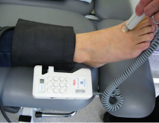
transducer : to measure the pressure we have at the compartment


Management : 

1. triage

2. if there is a cast or bandage, remove it immediately

**no compression

3. elevate the limb but not above heart treatment

4. emergency fasciotomy

2. Emergency Fasciotomy 

NF and GG


Necrotising Fasciitis vs Gangrene


** REMEMBER TO ALWAYS DO RESUSCITATION FIRST PRIOR OTHER MANAGEMENT (ABX, ANALGESICS, DM CONTROL)

Indication for amputation: 3D's

๐Ÿฃ Dying / diseased bone - PVD, infection, gangrene, trauma
๐Ÿฃ Dangerous - lethal sepsis, malignant
๐Ÿฃ Damn nuisance - gas gangrene, crush syndrome, severe malformation


Sample Xray


 Gas shadows seen extending from the foot up to upper calf 


Doppler 


Usually medical officer will ask for a doppler done and also ABSI. 
So try to get your doppler right. 
There are three spots that we must check : the Dorsalis Pedis Artery (DPA), Posterior Tibial Artery (PTA) and Brachial 

                   Normal ABSI: 1.0-1.3                                                    Normal wave: Triphasic
          ABSI 0.5-0.9: moderate to severe
ABSI >1.3: common in DM (may have severe calcifications)


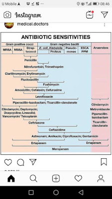
Antibiotics:

For NF: commonly polymicrobial bacteria can be seen. 
  • - IV Tazocin 4.5g QID (consist of piperacillin and tazobactem)
  • - IV clindamycin 600-900mg  TDS 
      • inhibitor of protein synthesis and protein  m synthesis 
      • effective for gram +ve
  • or Cefotaxime + metronidazole
 OTHERS:
  • - unasyn (beta lactam, inhibiting bacterial cell wall synthesis)
  • - ciprofloxacin : covers gram negative
Content Placeholder 4

Reference: 


Short terms;
- WD: wound debridement
- h/o : history of
- DM : Diabetes Mellitus
- DFU : Diabetes Foot Ulcer
- ABSI : Ankle Brachial Index

Tuesday, May 5, 2020

Orthopedic OSCE notes

 My Pocket Notes: Orthopedics OSCE


I always have this pocket notes in handy so I can study them without bringing the books along. They are useful when I need a quick glance and remember them quickly.
Especially the day before exam when we need to read a lot of information,
could not afford to waste any of those precious time for revision!


If you do not understand what I wrote about in the notes, you probably need to reflect and start studying the real books instead !


Friday, April 24, 2020

Test yourself - Pelvic Bones

Test yourself  - All about pelvic bonesss

You think you know it all? Try these and prove it !

Q1. Name the pelvic lines 


Answer:

A: Ilioischial line
B: Posterior rim of Acetabulum(ischio-acetabular line)
C: Shenton line
D: Iliopectinal line
E: Roof of Acetabulum
F: Anterior rim of Acetabulum (acetabulot-obturator line)
G: Shenton line
H: Tear Drop


Q2. Name the structure below


Answers:

A:  Right hemi-pelvis
B: Left hemi-pelvis
C: Sacroiliac joints
D: Sacrum
E: Pelvic Ring
F: Pubic Symphysis
G: Obturator Foramen

Q3. Name the structure and muscle attached to it


 Answers:

1. Iliac crest - abdominal muscles
2. Anterior Superior Iliac Spine : Sartorius muslce
3. Anterior Inferior Iliac Spine : Rectus femoris muscle
4. Greater Trochanter: Gluteus medius and minima
5. Lesser trochanter: Iliopsoas
6. Ischial Tuberosity: Hamstrings
7. Pubic Symphysis: Adductors

Other Notes:


 Muscles connected to the pelvic bones. 


This is a post specifically to help you revise what you know about the pelvic area, specifically for orthopedics. 

This is very important and popular to be asked in exam. 

I am sure you already know this structures from medical school, hope this exercise helps you to remember them again๐Ÿ˜

Good luck!


Tuesday, April 21, 2020

Unequal leg lengths : Limb Length Discrepancy

LLD : Limb Length Discrepancy


Limb Length Discrepancy (LLD)

Definition: where one limb is in a different length than its opposing limb, can be longer or shorter due to many causes.


Causes

1. Congenital (from birth)
2. Developmental:  can be from a childhood disease or injury that slows down or damages the growth plates
3. Post-traumatic
  • Car accident that causes a fracture which leads to shortening of the bone ends)

How to attend to patient?


Intro:


  • Confirm name, age
  • Ask chief complain and other important history (any motor vehicle accident before?)
  • Ask for consent and a chaperone if needed.
  • Always be friendly and make patient comfortable.

Inspection

For 4 aspects when the patient is Standing → check front, side and back↔ check for DAWSSS
  • Deformity
  • Asymmetry
  • Wasting
  • Swelling
  • Scar
  • Straight

Gait

Ask patient to walk normally and to walk on heels and toes.

Types of gait to look out for:

  • Short limb gait : dislocation of femur head or others -- confirm with bryant's triangle
  • Tredelenburg : Gluteus weakness, can be direct or indirect
  • Stiff knee : Osteoarthritis, Rheumatoid Arthritis
  • Antalgic gait : Pain, Limping and lag gait
  • High Stepping : Common among patient with peroneal nerve injury or palsy / sciatic nerve injury
  • Others: Spastic(Cerebral palsy), scissoring(UMN issue)

Supine on bed : Lie flat and inspect 

If there are gait issue go straight to finding the source

Check apparent length

- Measure the length from xiphisternum to medial malleolus of left and right
- Compare both sides

Check True length

- measure from Anterior Superior Iliac Spine(ASIS) to medial malleolus
-Compare both sides

Interpretation:
True shortening>Apparent shortening: Deformity compensated by pelvis tilt
True shortening<Apparent shortening: Fixed Deformity without any compensation


If there is one side longer than the other(suspect injury above knee joint) perform the specific tests

1. Galaezzi Test

  • Check for hip dislocation
  • Check when the patient is in supine position. 
  • Performed by flexing the knees while lying down so that the feet touch the surface and ankles at buttocks. 
  • Check the tibial length by flexing knee 90 degree and ankle 45 degree

Interpretation:
    • Shorter leg is the affected leg (femur head dislocated)
    • Knee shifts cephalad(outward) when the femoral length is reduced, and caudally(inward) when the tibial length is reduced
    • If it is parallel: LLD is below knee joint

2. Bryant's Triangle test


  • to check LLD above knee joint


Line A: Line connecting the greater trochanter of femur with ASIS
Line B: Vertical line done from ASIS towards the bed (perpendicular with bed)
Line C: Horizontal line starting from greater trochanter to meet end of line B
(compare length of line C on both sides for diagnosis)


Interpretation: 
    • diminution or increase in the length of line C : 
      • to measure supra trochanteric shortening
      • anterior or posterior displacement of the greater trochanter
      • can be cause by: dislocation of hip, fracture of femur neck , hip deformity, intra-trochanteric fracture
    • If both side are the same: the injury/ fracture might be in the middle of femur

Palpation and sensation


  • Tone - Hip Knee Ankle
  • Pain/tenderness - Hip, pubic symphysis, anterior superior iliac spine, axis, PSIS, greater trochanter
  • Temperature of each part. 

Motor Examination


  • Ideally would be to do a full motor examination, 
  • If the patient is in pain, focus on the examination specific to the patient's issue. They will be grateful that you help them attend to the issue ASAP. 
  • Range of movement for: hip, knee and ankle

Neurovascular Examination:


  • pulse
  • sensory
  • example: wriggle toes and see if they can feel which toe is moving

Other special tests: 


  • Thomas Test
    • Check the fixed and flexion deformity of hip
    • 1st picture is normal and 2nd is abnormal
    • Ensure their lower back is flat. 
    • Abnormal: Posterior thigh does not touch the table, knee flexion < 80 degrees

  • Tredelenburg test
    • examine strength of the abductor mechanism of hip
    • One legged stance, if the weight bearing hip is unstable the pelvis drop to unsupported side

  •  X-ray: check the AP view and lateral view


Ideally to take full history, examine spine and knee, access patient with radiography.

Other references:
1. http://www.limblength.org/conditions/limb-length-discrepancy-lld/
2. https://www.slideshare.net/ManishShetty8/clinical-examination-of-hip-122918037