Showing posts with label house officer. Show all posts

Saturday, July 23, 2022

Paediatric as HO

Paediatric books to study in HO

In our hospital when I am in paediatric department posting, mainly using the paediatric protocol book. It is quite important as we use it when we have presentations in department. 

Other than paed protocol, I found that the following other books and website is also useful. 


Neonate Paediatric / Oncology paediatric:

Paediatric Protocol

MOH Paediatric Protocols for Malaysian Hospitals, 4th edition


General Paediatric:

 Ampang guideline

https://myhow.files.wordpress.com/2013/11/hoguidepeds1.pdf

Physical examination:

MRCPH website

https://mrcpch.paediatrics.co.uk/

Medications

Frank Shan book
**highly recommend to get the physical copy, as some alignment in the ebook is not as accurate. 


Things to prepare:

  • Pens
  • Measuring tape
  • Mini calculator (either those pocket size or the ones that you can hang on your landyard)
  • Scissors
  • Durapore

Notes to bring along

  • Fluid requirements 
  • Growth development
  • Growth chart
  • Normal readings of blood test
  • Normal values for vital signs (RR, BP percentile) 
    •  it is quite challenging and difficult to memorise the whole chart, easier to bring it around and refer when needed. 
  • mini toy/ cute things 
    • (to help kids to calm down before any procedures, eg: taking blood/ punctures)
    • I have a mini duck with me most of the time, and i find it very useful in making the kids happy and cooperate. However later on I realise they just like anything that is in bright colour, not necessarily toys.  Even a cute plaster could make them feel so happy. 

Good Attitude

  • kids are often affected by our mood/ emotions, so do make sure you are treating them with joy, patience and love.
* Always stay vigilant, observant and a good attitude helps you get through paediatric posting. 
You might enjoy it in the end too!


Sunday, July 10, 2022

Abdominal examination for paeds

Abdominal examination:


General Examination

  • General surroundings
    • equipments : NG tube/ PEG tube
    • nutrition: milk, food
    • Growth chart
  • Hand
    • finger clubbing
    • crt, pulse volume, warm/cold peripheries
    • rashes? 
  • Face
    • eyes: anemia? edema?
    • mouth: gum bleed/ hydration
    • cyanosis
    • overall inspection
  • Neck
    • spider naevi
    • inflammation/ edema on the neck
    • lymph node
  • Abdomen
    • Inspection
      • shape
      • distension
      • masses
      • rashes/scar/striae/stoma 
    • Palpation
      • pt should be in supine position, gently ask if it is comfortable to lie down if having abdominal distension
      • superficial, deep palpation
      • liver/ spleen/ kidney palpation/ abdominal mass
    • Percussion
      • shifting dullness/ fluid thrill
    • Auscultation
      • resonance/ dull
  • Lower back
    • sacral edema?
  • Lower limbs
    • pedal edema
Sample video


Special cases
  • Nephrotic syndrome : ascites/ periorbital edema/ pedal edema/ sacral adema/ pleural effusion

Causes of hepatomegaly

  • 1.  Structural: Extrahepatic biliary atresia, choledochal cyst, intrahepatic biliary hypoplasia, congenital hepatic fibrosis, polycystic disease

    2 . Storage/ metabolic:

      – Carbohydrate- glycogen storage disease (Type 1,3,4,6), hereditary fructose intolerance, galactosaemia,, mucopolysaccaridoses-

    – Mineral: Wilson’s disease, juvenile haemachromatoses

    -Nutrition: Protein calorie malnutrition, TPN-Bile flow: progressive familial intrahepatic cholestaisis syndrome

    -Protease: Alpha-1-antitripsin

    -Electrolyte: CF

    – Amino acid: Tyrosinaemia type 1, urea cycle disorder

    -Lipid – Gaucher, Nieman-Pick diease, cholesteryl ester storage disease

    3. Haematological: Thalassaemia, sickle cell disease, ALL, AML CML

    4. Heart/ Vascular: Congestive heart failure, constrictive pericarditis, obstructive IVC, Budd-Chiari syndrome

    5. Infection: Viral – rubella, CMV, coxsackie virus, echovirus, hepatitis ABCDE, EBV  ; Bacteria – E.coli TI, TB, syphilis ; Parasite – Malaria, toxoplasmosis, Schistosomiasis


Reference: 

Friday, December 17, 2021

Fat embolism

Definition: 

Fat emboli:

- a complication of trauma/ surgery that involve intrumentation of femoral intramedullary canal

- it is a response/ manisfestation of fat globules that may enter the blood stream

Fat embolism:

- a process by which fat emboli passes the bloodstream and lodges within the blood vessel

Fat embolism syndrome:

- serious manisfestation of fat embolism that causes multi system dysfunction


Causes: 

1. mechanical theory

- fat droplets from bone marrow enters the vessel

- increase of intramedullary pressure and cause fat/marrow to enter the bloodstream. which later could lodge in the lungs as emboli

- causes inflammation and local ischemia

2. metabolic theory

- stress from trauma that causes change to the chylomicron that causes formation of fat emboli

or

1. trauma related

- fracture at long bones: especially femur fractures

2. non trauma related

- liver disease, shock, bone tumor lysis


Symptoms:

- asymptomatic within the first 12-48 hours

Gurd's criteria: 2major 1 minor or 1major 4 minor

  • Major: 

- hypoxemia, petechial rashes, neurological symptoms, pulmonary edema

  • Minor: 

-tachycardia, fever, retinal changes/ renal changes/ fat macroglobinemia, jaundice, 

- drop in Hb, increase ESR, thrombocytopenia


Investigation: 

- FBC

- ABG

- RP/LFT

- CXR : ground glass appearance / snow storm appearance


Management:

- stabilise the patient

  • Airway : no obstruction
  • Breathing: oxygen support
  • Circulation: two large branulla with fluid support 
    • hemodynamically: Hb? any blood loss
    • hydration: 3L/d

1. monitor vital signs : BP, PR, SPO2, temperature

2. inform 

- MO incharge, anaest (ventilator support)

- keep in view the need of doing CT brain to exclude other causes

3. rigid fixation of fracture within 24 hours

4. diagnosis of exclusion

5. DVT prophylaxis 

6. stress ulcer prophylaxis


Reference: 

1. https://www.orthobullets.com/basic-science/9055/fat-embolism-syndrome

Compartment syndrome

Definition:

a rise in pressure within a closed osteofascial compartment, resulting in impaired local circulation compartment. 


Causes:

high risk injury:

- fracture that causes inflammation to the muscle where it has no place to expand therefore compressing on the vascular structure and nerves

- operation

- infection

- trauma: crush injury

- burn injury

- tight plaster cast


clinical features

5P s

  • PAIN: pt will complain excrutiating pain, just like they wanted to have their legs cut off
  • Pallor
  • Parasthesia
  • Pulselessness
  • Paralysis

Passive stretch test: positive 

- can be seen in the video below 2:50 onwards


transducer : to measure the pressure we have at the compartment


Management : 

1. triage

2. if there is a cast or bandage, remove it immediately

**no compression

3. elevate the limb but not above heart treatment

4. emergency fasciotomy

2. Emergency Fasciotomy 

Friday, November 5, 2021

Fluid and Resus common QnA for HO

Part 1: Fluid and its components

Q1. What are the common cause of fluid loss in surgery?

  • apparent loss: diarrhea, vomiting and high output stoma
  • 3rd space loss: 
    • loss of water, electrolyte and colloid particles into interstitial space
    • which could contribute to edema
    • Intestinal obstruction, pancreatitis and ascites
  • others: insensible fluid loss (hyperventilation/pyrexia), stress response

Q1a. How patient loss fluids from IO?

  • apparent loss: vomiting
  • 3rd space loss: 
    • increased secretions
      • bowel obstruction will cause bowel to secrete a lot of secretion to overcome the obstruction. 
    • mucosal edema, so fluid not absorbed
      • so there will be a lot of accumulation of fluid that leads to third space loss
      • fluid accumulation in bowel can reach up to 6L
      • that could lead to hypovolemic shock

Q1b. How patient loss fluid from pancreatitis?

  • systemic inflammation 
    • inflammation causing release of inflammatory cytokine and other pro-inflammatory mediators,
    • leading to capillary leakage
    • thus loss of circulatory albumin and fluids to interstitium
      • capillary leakage cause fluid shift to third space and then hypotension leads to hypovolemic shock.

Q2. What is the main difference between crystalloid and colloid?

  • molecular size
    • affects shifting of fluid where low molecular size - low tonicity


Q3. How does fluid moves in human body?

  • from low concentration to high concentration 
    • big molecules fluid (colloids) 
    • has ability to pull fluids from other compartments : oncotic pressure
  • opposite of oncotic pressure: hydrostatic pressure
** not through pressure gradient / osmosis

Q4. What is isotonic?

  • a solution concentration that is similar to plasma
  • the osmolarity of plasma is around 300 mosm/L
    • Normal saline: 308
    • Half saline: 154 (hypotonic)
    • 3% saline: 1026 (hypertonic)

Q5. What is the difference between Normal Saline (NS) and Hartmann(HM) solution?

  • Hartmann has additional potassium, lactate, calcium
  • the most "physiological" solution

Q5a. What is the function of lactate in HM

  • lactate will be metabolised by liver to HCO3, thus acting as buffer
    • especially in met acidosis

Q5b. Can we use HM as resuscitation fluid?

  • No
    • usually when patient needs resuscitation, already with multiple organ failure or impairment
    • so although Hartmann has lactate that could help as buffer, the liver is unable to convert lactate to bicarbonate, which could lead to accumulation
    • the accumulation of lactate will worsen the metabolic acidosis
    • and Pt in acidosis usually has hyperkalemia, Hartment contains K+ which would further worsen the situation


Q5b. So when do we use hartmann?

  • as maintanence, especially in those who need replace electrolyte loss (diarrhea and vomiting)

Q6. What is the function of Dextrose 5%?

  • provide hydration
  • the have glucose in solution not for calorie to avoid lysis and avoid hypotonic
    • calorie in D5: 170/L
  • it is just to render solution isotonic once infused in the circulation, once they reach liver will convert into free fluid
    • - provide free water that can pass through membrane pores, expanding both intracellular and extracellular spaces


Part 2: Fluid and resuscitations

Q7. 60year old, 70kg man presents with diarrhea and vomiting for 1 week. brought in with hypovolemic shock
Outline your management for this man

- ABC
  • assess airway
  • breathing
  • circulation
    • check the vital signs: unstable/ stable
- insert 2 large bore needles and give IV NS
- run fast 1 pint NS

Q7a : If patient doesn't respond to fluid resus? 

  • reaccess: if the volume is improved, but patient still hypotensive, he might have other component of shock
    • for example: septicaemic shock --> we might need to start inotrope for vasoconstriction
    • if cardiogenic shock / has underlying IHD --> get an ECG, and we might need to start with dopamine or dobutamine as the inotropic support. 
  • if volume is still low, 
    • can infuse COLLOID to hold the fluid in the circulation
    • colloid has oncotic pressure that will hold the fluid intravascularly, thus maintain the BP
  • So why we cant give colloid straight away for resus?
    • Colloid causes shifting of fluid out of the cell, worsen the hypoperfusion
    • in shock, circulation fails and tissue is hypoperfused, if we infuse hypertonic solution all fluid will move from the tissue into the intravascular system . 
    • Therefore load with volume first (crystalloid)
      • resume the circulation
      • let them reach the heart, brain and kidney
      • after that infuse colloid to hold the volume. 

Q8: Define shock

  • must have 2 components
    • circulatory failure: seen via vital signs
    • inadequate tissue perfusion : seen via low SPO2
      • sequelae of low perfusion
      • multiorgan failure

Q9: Why we cant have central line when patient is in shock?

  • it is about the catheter's caliber. the shorter the calibre, faster the infusion. 
    • if central line, it has long calibre and the rate of infusion is slower
    • insertion takes a lot of time
  • in shock we need large supply of fluid for the patient 
    • Poiseuille law

Q10: How do you know patient responded to your fluid resuscitation? what are your AIMS?

  • vital signs 
    • HR <90
    • BP >90/60 , MAP >60
    • SPO2 >95%
    • RR <20
    • u/o  >0.5ml/kg/hr


Saturday, June 26, 2021

OBGYN books for Houseman (HO)

Books recommended 
while having placement 
in Obstetric & Gynaecology 
as a (HO/houseman) 
in Malaysia

1. Clinical protocols in OBGYN for Malaysian
2. CPG Malaysia
3. HUSM labour suite - by Prof Adibah
4. Ampang notes O&G guideline
5. Quick management for gynae - by Dr Lee Say Fatt
6. Medicorp
7. Others


Protocols for OBGYN (Msia)
I find that it is a good guide for me when i am working OBGYN Houseman. A good book for those who practice as a doctor for hospitals in malaysia

Sunday, May 2, 2021

Pulmonary Embolism

 Pulmonary embolism (PE)

it is not complete but do include key points that we need to know to work in ward. 

pathophysio: 

  • it results from DVT - venous thromboembolism
  • venous stasis, endothelial injury and hypercoagulability. 


bridging:

  • egfr>30: s/c clexane BD
  • egFR 15-30 - s/c clexane OD
  • eGFR <15 - IVI heparin (kena monitor coag 6 hourly)
  • Warfarin affects the APTT & PT value
  • Clexane - APTT (to help INR reaches the aim faster)

  • Aim INR 2-3, once INR >2, off clexane, bridging complete
1st to 3rd days for warfarin just trial with dose 5mg/5mg/3mg, 
4th day start adjusting dose base on INR

- pharmacist has special counsellor or booklet for the patient to understand the use of warfarin. with constant follow up and also diet suggestions (no green leafy veges)


BOOKLET LINK: for BM, Eng, Chinese

Investigations(Ix):

1. ECG :
look for ST changes S1Q3T3

- deep S wave in lead I, Q wave in lead 3, T inversion in lead 3
- ST depression, RBBB
- P. pulmonale


2. blood Ix: trop t and ck stat

3. basic supportive tx: HFM, transfer to acute bed

request for :

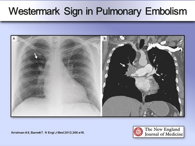
- CXR : hamptoms hump: wedgemark sign, westermark sign: pulmonary oligaemia in the affected segment


- CTPA : thrombi within pulmonary

https://epos.myesr.org/poster/esr/ecr2020/C-04154

- V/Q scan:
  • for those who have clot in the past, and have a previous episode before, or those who has acute kidney damage/ckd.
  • clear cxr
- ultrasound for lower limb: to rule out lower limb DVT due to long sitting/surgery...

- start DAPT

4. echo:
- look for RV dilation and hypokinesia. - acute rv failure--> increase afterload, rv cannot unload sufficiently causing rv dilated. impinge on LV cause LV to decrease output and supply to CA
- it increases o2 demand which CA could not meet, results in ischemia and necrosis

MASSIVE PE:
HYPOTENSION BP<50 for at least >15MINUTES, HR <40 - usually with evidence of MI and hyocardial dysfunction

Well's score for PE

low<2, mid 2-6, high >6
1. clinical signs of DVT -3
2. previous surgery / long traveeling>4hrs -1.5
3. previous DVT and PE- 1.5
4. HR>100 - 1.5
5. other diagnosis seem less likely -3
6.hemoptysis -1
7. cancer -1


Management (Mx):

anticoagulant
- without hypotension: LWMH, fonda/ DOACS
- unstable/ bleeding: parental anticoag 5-10d, switch to warfarin overlap 3-7d before discontinue heparin when INR >2 for >2d
- continue anticoag for 3-6months
thrombolytic therapy:
STK 25mu if pt high risk massive PE/ with cardiopulmonary arrest.
other mx: 
1. rivaroxaban 15mg BD for 21d then 20mg OD
2. dabigatran (expensive)

Thursday, April 8, 2021

Common Investigations

 Special workups / short terms for the investigations needed: 


IJC exchange

  • One IJC
  • General set
  • Syringe 10cc x2
  • Sterile prep
  • 5cc syinge x2
  • Lignocaine
  • Gown, apron
  • Hep saline(blue) in 100cc normal saline
  • Blue sheet
  • Sterile gloves
  • Blue needle x1
  • Green needle x1
  • One set ijc rm240 must make sure correct ijc - 2flo or 3 flo
  • Suture set, syringe(blue) and need.


Chest tube insertion template

Retrospective entry


Upon primary survey noted patient is having right tension pneumothorax evidence by: hyperresonance, reduce air entry over affected site and trachea deviated to opposite site.


Decided for chest tube for definite treatment after immediate needle thorachocenthesis.


Chest tube was inserted by_____.

Explained to patient regarding the need and possible complication of chest tube insertion; patients understood & agreed.


Chest tube inserted at safety triangle with open method, using chest tube size ___, and anchored at ____cm


Post insertion, chest tube functioning evidence by gushed of air.


No immediate complication observed.


Plan;

-Keep patient propt up patient 30-45 degree

-for cxr post chest tube insertion

-keep high flow mask

-keep spo2 more than 95%

-to teach patient for lung exercise.


Tuesday, March 16, 2021

drug doses to know in medical


LIST of important drugs and doses

 IV hydrocot 200mg stat

S/c Fondaparinox 2.5mg OD/7.5mg OD

S/c Heparin 5000u BD/7500u BD

S/c Morphine 5mg/7.5mg TDS

S/c Clexane 40mg/60mg OD

T Ranitidine 150mg BD

IV Ranitidine 50mg TDS

Mist calminative 15mls TDS

Mist Na Citrate 15mls/30mls OD

T EES 400mg BD

T Zinnat 250mg BD

T Ponstan 500mg TDS

T Voltaren  50mg TDS

T Papase 11/11 TDS

T Gelusil 11/11 TDS

IV Maxolon 10mg TDS

C Celebrex 200mg BD /400mg OD

C Tramal 50mg/75mg TDS 

T Arcoxia 90mg OD

Syp MMT 15mls TDS

Syp Benadryl 15mls TDS

T Piriton 4mg/8mg TDS

IV hydrocortisone 200mg stat, 100mg QID

T Cilostazol (claudication pain) 100mg OD

T Aspirin 150mg OD/300mg STAT

IM nubain 20mg stat

T/IV Bisolvon 8mg TDS 

T Labetolol 100mg TDS 

T Adalat 10mg TDS

T Methyldopa 250mg TDS

Ural sachet 1/1 BD/TDS 

IV/T lasix 20/40/80 mg BD

IM/IV Pethidine 50mg

T Rocaltriol 0.25 mcg OD

T calcium lactate 600mg BD

T levenogestrel 1.5mg stat

Inj Ertapenem 1gm OD

Inj Piptaz 3.375g 

T/IV Tranexamic acid 500-1000mg TDS

IV Tramal 50 mg/75mg TDS

IV Maxolon 10 mg TDS

IV Cefuroxime 1.5 stat, 750mg TDS

IV Unasyn 1.5g STAT and TDS

IV Cefobid 2g stat, 1g BD

IV Ceftriaxone 2g stat, 2g/1g OD

IV Cefepime 2g stat, 1g BD

IV Meropenem 500mg tds/qid

IV Imipenem 500mg tds/qid

IV Ampicillin 2gm Stat 1gm QID

IV Cloxacillin 1g STAT, 1g/500mg QID

IV Flagyl 500mg TDS

IV Augmentin 1.2g STAT, TDS

T Augmentin 625 mg BD

T Unasyn 375mg BD

T Doxycycline 200mg stat, 100mg bd

T Cloxacillin 500mg QID

T Azithromycin 500mg OD

T Flagyl 400mg TDS

C Tramal 50mg/75mg TDS

T Maxolon 10mg TDS

T Doxazosin 4mg OD 

T Finasteride 5 mg OD

T Cardura 4mg ON

T Hytrin 4mg ON

T. Metoprolol 50mg BD

IV vancomycin 500mg QID/1g BD over 1hr40min - MRSA  

Cream chlophelamine 

T. Cetrizine 10mg stat and OD

Monday, March 8, 2021

HO medical - EMERGENCY management

Some of the notes seniors passed down which is very useful. I edited and add in some points i get during my posting as well. 

Big thumbs up to them who prepared the list and Thank You💕💖

  1. Hypokalemia
  2. Hyperkalemia
  3. Hypoglycemia
  4. Hyperglycemia
  5. Chest Pain
  6. Hypotension
  7. Asystole
  8. VT
  9. Atrial Flutter
  10. SVT
  11. Fit
  12. GCS drops
  13. Aggressive behavior
  14. Nausea/ vomiting
  15. Diarrhea
  16. Hematemesis
  17. Shortness of Breath
  18. Other electrolytes
  19. UGIB
  20. Anaphylaxis
  21. Dengue


1. Hypokalemia (K+ <2.5)

- ECG STAT to look for hypokalaemic changes, check whether patient symptomatic.

- 1g KCL in 100cc NS in 1 hour

                or

    2g KCL in 200cc NS over 2hour (according to K+ level)

- with continuous cardiac monitoring

- Add KCl in drip (if patient on drip), mist kcl 15mls tds OR T. slow K 600mg/1.2g OD

- Check TFT, RP, VBG, UFEME

- Monitor v/s

- ECG (any ecg changes)

- Inform MO stat if symptomatic

- Off potassium supplements once K+ >4

- Repeat RP 1 hour post correction

- Daily RP until K+ stable


 2. Hyperkalemia (K+ >5.2)

- ECG STAT to look for hyperkalaemic changes, check whether patient symptomatic

- Off potassium supplements

- To serve lytic cocktail 

(10cc of 10% calcium gluconate in 10 minutes - slow bolus with cardiac monitoring + 50cc D50% glucose + 10 unit actrapid)

- If still high, repeat lytic cocktail 

Peritoneal dialysis?? Haemodialysis??

- T. kalimate 5-10g TDS

- Off kalimate once K+ <5

- Repeat RP 1 hour post correction

- Monitor RP daily till stable


3. Hypoglycemia (Reflo <4)

- Omit insulin

- Encourage patient to take orally (sweets, bread with jem, milo)

- If too low (<3.5) with symptoms, give 20-50cc D50% then repeat reflo 30mins

- If reflo 4-6, half the dose of next insulin


4. Hyperglycemia (Reflo >12)

- Serve S/C 6-10 unit actrapid STAT (depends on reflo) if patient not on insulin/insulin served earlier on.

- If patient already on insulin and not served yet, to serve usual dose.

- If persistently high despite insulin to start top up regime based on BMI. Take VBG, lactate and urine ketone dipstick TRO DKA.


5. Chest pain

- ECG STAT.

- If suspected ST elevation, to inform MO for referral to cardio KIV thrombolyse.

- S/L GTN, maximum 3 times. If persistent pain to start IV morphine with IV maxolon. If pain persists despite morphine to start IVI GTN.

- Oxygen

- Aspirin crush 300mg, Plavix 75mg

- Take cardiac enzymes(CK) and Trop T as baseline, to repeat at 6 hours later with ECG


6. Hypotension

- Determine cause (Septic, hypovolaemia, cardiogenic etc)

- Repeat manual BPx2 first to confirm

- Run 1 pint (250cc) NS fast 15-30minutes

     if no contraindication (watchout if patient have ROF).

     If still low, for another try.

- Inform MO is persistently low, KIV start IVI noradrenaline and adjust accordingly

- Regular BP monitoring (ideally every 15mins)

- KIV Gelafundin (colloid) @ inotrope

- Grey branula at neck/femoral line

- IV Noradrenaline 0.2mcg/kg/min

- KIV add another intropes if low despite high dose 1st intropes


7. Asystole

- Inform MO and call CRASH 

- Prepare Resus trolley and intubation kit

- Manual bagging 15L/m (even for ventilated patient)

- Straighten bed and commence CPR

- Transfer to acute bay

- Vital signs, reflo and cardiac monitoring

- Insert 2 large bore branula at least green at big veins (femoral/neck/cub fossa) and take all routine bloods including ABG, reflo and cardiac enzymes (run urgently)

- Run 1 pint NS fast if no contraindications

- Prepare IV adrenaline (1mg every 5 mins 3 cycles)

                IV Atropine 1mg every 3-5 mins (3x)

- If patient survive,

  •     take ABG post intubation,
  •     ECG,
  •     septic workup (if infection is suspected),
  •     D dimer (if PE is suspected)

- Keep BP >90/60, MAP >60.

- Insert CBD and strict IO monitoring (Keep U/O > 30cc/hr)

- Start inotropes if low BP

- IV panto 40mg OD to prevent gastric ulcer

- Insert Ryles tube, KIV start feeding later (refer to dietitian)


8. VT

- Inform MO

- Prepare resus trolley

- Vital signs and continuous cardiac monitoring

- If pulseless, for defib and 5 cycles of CPR. Repeat if unsuccessful.

- If pulse present but hemodynamically unstable, for urgent cardioversion and IV

lignocaine

- If pulse present, for IV amiodarone


9. Atrial flutter

- Inform mo

- Vital signs and continuous cardiac monitoring

- If hemodynamically unstable, for urgent DC shock and rate control meds.


9a. Fast AF

- Nasal prong O2 (NpO2)

-Continuous cardiac monitoring

- IV Digoxin (if HF) 0.25mg every 2hr, up to 1.5g within 24hr

- Metoprolol 25mg/100mg BD (absence of HF)

- KIV IVI Amiodarone 300mg over 30mins

- KIV cardioversion if hemodynamic not stable


>10. SVT

- Inform MO stat

- Vital signs monitoring and continuous cardiac monitoring

- If hemodynamically unstable, for cardioversion

- If stable for carotid massage (if no bruit) dan valsalva maneuver

- If persistent, need to give IV adenosine 6mg and flush with 20cc NS.

  • then 12mg then 12mg
  • if not reverted, IVI amiodarone 300mg over 30 minutes

Contraindicated for Asthma patient.

  • Second bolus 12mg can be given after 5 mins with NS flush.

- If persist consider another drug (verapamil etc)


11. Fit

- Remove patient from dangerous objects

- Prepare IV diazepam 5mg STAT, if persist repeat dosage (max 3 doses with 10 mins gap)

- Put on HFM 10-15L/min

- Vital signs monitoring

- Put patient on left lateral position

- If fit persist, KIV IV phenytoin (loading and maintenance)

- Repeat bloods including electrolytes, KIV for CT brain/ LP

- If resolve, continue vitals monitoring, fit charting, - GCS charting


12. GCS drops

- Vital signs and reflo

- Inform mo

- Transfer to acute bay

- Refer CRASH KIV for intubation

- Determine cause

- Insert line and repeat bloods including ABG, septic workup Request for CT brain

-Treat according to cause


13. Aggressive behaviour

- Approach calmly

- Ask help from security guards or male staff

- IV haloperidol 5mg STAT

- 4 points restraints

- Repeat bloods TRO causes

- Refer psych if persistent aggressive behaviour


14. Nausea/vomiting

- IV maxolon 10mg STAT

- Start ORS

- If significant loss, start IVD maintenance

- Monitor RP

- Find the cause


15. Diarrhea

- ORS per purge

- KIV lomotil

- If significant loss, start IVD maintenance

- Monitor RP

- Find the cause


16. Hematemesis

- Inform MO

- Rule out UGIB (inspect vomitus)

- PR examination to look for melena

- Vital signs (look for compensated or decompensated shock)

- If significant blood loss to insert 2 large bore branula and repeat bloods

- IV tranexamic acid 1g STAT and 500mg TDS, IVI pantoprazole 8mg/hr.


17. SOB

- Examine lungs, check vitals

- Determine cause, repeat CXR if needed

- Refer CRASH if necessary (SpO2 unable to maintain with oxygen

supplementation or clinically worsening SOB)

- If intubated, check if ETT dislodged or too deep (if yes, to readjust)

- Start ocygen supplementation (according to SPO2 and clinical). If known case of

COPD to start VM.

- Watchout for respi distress, keep SpO2 >95%

- If significant SOB with Sp02 drop, take ABG, FBC (TRO anaemia), Cardiac

enzymes, Trop I (ACS is suspected), D dimer (TRO PE)

- If ronchi (asthma) - Neb AVN STAT, IV hydrocortisone 200mg STAT (if moderate

to severe). Reassess post neb, if worsening can try back to back neb.

- If PE is suspected, to take D- dimer KIV CTPA. Chest referral, KIV start clexane

- If pneumonia (HAP/aspiration) is suspected, to take septic workup and CXR and

start antibiotics.

- If fluid overload, to serve IV frusemide 20mg STAT (to check BP before serving),

adjust IVD and fluids intake

- If pleural effusion, KIV for tapping


>18. Other electrolyte deranged management


  • Hypomagnesaemia:
    • IVI MgSO4 1 ampule in 100cc NS over 1 hour
  • Hypocalcaemia:
    • - ECG
    • - IVI CaCO3 1 ampule in 100cc NS 
    • over 1 hour @
    • - IVI Calcium Gluconate 1 ampule in 
    • 100cc NS over 4 hours
    • * Mild: Tab CaCO3 500mg BD/TDS

  • Hypophosphatemia:
    • - IVI KH2PO4 1 ampule in 100cc NS 
    • over 4 hours

  • Hyponatremia:
    • - If no ROF, give IV drip NS 3-4  pints/24 hour


19. Upper GI Bleeding:

- 2 large bore branula 

- Run fluid 

- FBC, Coag profile, GXM 4pint blood

- IV Pantoprazole (PPI) 80mg stat 

and IVI 8mg/hr

- Inform MO


20. Anaphylaxis:

- IV Hydrocortisone 200mg

- IV Piriton 10mg

- IV Maxolon 10mg stat & TDS

- Oxygen 

- IV fluid

- If BP drop,IV/IM Adrenaline 1:1000


21. Dengue1:

- Dengue fever day __, __ warning signs, in __ phase (if defervescence phase point taken at__˚C), V/S, on drip __cc/kg/H, latest FBC reviewed. -

- Next FBC at __am/pm, cont drip __cc/kg/h


* WARNING SIGN: 

- Tender liver - Abdominal pain

- Mucosal bleed

- Persistent vomiting ≥3x + diarrhea  ≥3x/24 hr 

- Fluid accumulation(ascites/pleural E)

- Restlessness/altered conscious level

- Inc haematocrit, reduced platelet


Dengue2:

- N haematocrit: Male: >45; Female: >40

- Raised haematocrit in active smoker & obese pt is normal

- If pt took PCM, take Temp > 6hours after that, to count defervescence phase (< 38˚C)

- Dengue IgM & IgG positive high risk DHF (If systemic bleeding, give IV Traxenamic acid 500mg TDS)

- Ideal Body Weight in Dengue: 

Male: (Ht-152.4) x 0.91 + 50

Female: (Ht – 152.4) x 0.91 + 45