Showing posts with label clinical skills. Show all posts

Sunday, July 10, 2022

Abdominal examination for paeds

Abdominal examination:


General Examination

  • General surroundings
    • equipments : NG tube/ PEG tube
    • nutrition: milk, food
    • Growth chart
  • Hand
    • finger clubbing
    • crt, pulse volume, warm/cold peripheries
    • rashes? 
  • Face
    • eyes: anemia? edema?
    • mouth: gum bleed/ hydration
    • cyanosis
    • overall inspection
  • Neck
    • spider naevi
    • inflammation/ edema on the neck
    • lymph node
  • Abdomen
    • Inspection
      • shape
      • distension
      • masses
      • rashes/scar/striae/stoma 
    • Palpation
      • pt should be in supine position, gently ask if it is comfortable to lie down if having abdominal distension
      • superficial, deep palpation
      • liver/ spleen/ kidney palpation/ abdominal mass
    • Percussion
      • shifting dullness/ fluid thrill
    • Auscultation
      • resonance/ dull
  • Lower back
    • sacral edema?
  • Lower limbs
    • pedal edema
Sample video


Special cases
  • Nephrotic syndrome : ascites/ periorbital edema/ pedal edema/ sacral adema/ pleural effusion

Causes of hepatomegaly

  • 1.  Structural: Extrahepatic biliary atresia, choledochal cyst, intrahepatic biliary hypoplasia, congenital hepatic fibrosis, polycystic disease

    2 . Storage/ metabolic:

      – Carbohydrate- glycogen storage disease (Type 1,3,4,6), hereditary fructose intolerance, galactosaemia,, mucopolysaccaridoses-

    – Mineral: Wilson’s disease, juvenile haemachromatoses

    -Nutrition: Protein calorie malnutrition, TPN-Bile flow: progressive familial intrahepatic cholestaisis syndrome

    -Protease: Alpha-1-antitripsin

    -Electrolyte: CF

    – Amino acid: Tyrosinaemia type 1, urea cycle disorder

    -Lipid – Gaucher, Nieman-Pick diease, cholesteryl ester storage disease

    3. Haematological: Thalassaemia, sickle cell disease, ALL, AML CML

    4. Heart/ Vascular: Congestive heart failure, constrictive pericarditis, obstructive IVC, Budd-Chiari syndrome

    5. Infection: Viral – rubella, CMV, coxsackie virus, echovirus, hepatitis ABCDE, EBV  ; Bacteria – E.coli TI, TB, syphilis ; Parasite – Malaria, toxoplasmosis, Schistosomiasis


Reference: 

Saturday, November 6, 2021

Electrolyte Imbalance QnA for HO

 House Officers must know in ward 

- Electrolyte imbalance


Q1: 50year old, 70 kg, male with persistent diarrhea and vomiting for 3days due to food poisoning. He is dehydrated. Vitals are stable, looks lethargic. Na: 128mmol/L

a. How to correct his hyponatremia

    - calculate first
  • Na deficit (mmol) : (desired Na level - serum Na) x bw x fraction
  • 0.6 x kg x (desired Na - serum Na)
    • desired Na :  135-145
    • Na deficit is 0.6 because extracellular comprises 60%. 
    • If its potassium (intracellular) therefore 0.4 cause it is 40%
  • 0.6 x 70 x (135-128) = 294mmol/L
    - to add with daily requirement
  • infusion = deficit + requirement
    • requirement: 1-2mmol/kg/d (we usually use the lower range when calculating)
  • infusion: 294 + 70 = 364 mmol/L
- then how do we deliver 364mmol/L to pt?
  • 1 pint NS = 77mmol/ L
  • 1L NS = 154mmol/L
  • 364 / 77 = 4.7 pints, 
  • therefore 5 pints of normal saline over 24 hours is needed.

b. the next day his repeated Na is normal: 136, 

how do you plan for fluid regime ? for maintenance?

  • normally a person need 30-40ml/kg/d (fluid requirement)
    • general normal weight for man: 70kg,  normal weight for woman : 60kg
  • if we take the middle value fluid requirement (35ml/kg/d), 
    • the man will need 2450ml (around 5 pint as well)
    • women is 2100ml, around 4pint. 
thats why usually we see usually 
  • 5 pint (2/3 pint NS + 2/3 pint D5%) given to man, 
  • 4 pint(2 pint NS + 2 pint D5%) given to women. 
** WHY 2 or 3 pint NS? for example a 70kg man we take 140mmol Na requirement, 1 pint NS is 77mmol, therefore the man only need 2 pint NS. but his fluid requirement is 5 pints a day, that's why we need to add another 3 pint D5 to complete his daily fluid requirement. 


Q2. what should you watch out during sodium correction?

  • Na correction should not exceed 10mmol/L/24 hours
    • thats why we use the lower normal when correcting sodium
  • even in severe hyponatremia, if Na only 120, your aim of correction is only up to 130, and not up to 135 in 24 hours. 
  • if there are hypernatremia (rare), mostly we try to find out the associated conditions/ related underlying issues. usually will be given diuretics to excrete excess sodium. 


Q3: 50year old, 70 kg, male with persistent diarrhea and vomiting for 3days due to food poisoning. looks lethargic, no ecg changes.  K: 3.2mmol/L

a. how to correct his potassium?

  • Deficit = (desired K - measured) x Body Weight (BW) x 0.4
    • (3.5-3.2) x 70 x 0.4 = 8.4mmol
    • 70 for maintanence , 
    • maintanence + deficit = infusion
      • 70+8.4 = 78.4
    • 78.4/13.3 = 5.8gm
      • we take normal daily requrement of K : 1mmol/kg/d, therefore we need to convert into gm  by dividing 13.3
      • 1gm = 13.3mmol
    • so we need to correct at least 5 gm first
      • can correct with 1g KCL in each pint NS (total 5gm)/24hours

b. what is the maximum/ safe dilution of K+ in 1 pint of NS?

  • dilution cannot exceed 40mmol/L
  • which also means cannot exceed 20mmol/pint = 20/13.3 = 1.5gm KCl
  • we cannot give more than 1.5gm KCl in a pint of NS

c. what is the safe titration of K+?

  • titration of potassium cannot exceed 10mmol / hour
  • if we need fast correction due to symptomatic, 
    • max is 1gm KCl in 100cc NS over 1 hour, not faster than that
  • If need 2gm infusion, it must be in 2 hours.
    • never bolus correction, must always use safe titration
    • 2gm KCl infusion in 200cc NS over 2 hours
  • OTHERWISE can cause CARDIAC ARREST! 

Q4: How do we know the patient has hyperkalemia?

  • Cut off point for hyper K: 
    • K+ > 5.5
    • symptomatic ECG changes: tall tented T waves, absence of P wave, broad QRS, PR prolong
    • it became dangerous when the ECG changes go towards heart block type of ECG with tall tented T wave --> severe hyperkalemia --> lead to arrythmia then asystole

Q5. what is the fx of each components in lytic coctail? 

  • lytic cocktail: treatment for hyperkalemia
  • Calcium gluconate - 10ml 10% calcium gluconate
    • for cardio protection
  • Insulin / actrapid 10u : 
    • drive K+ into cell together with glucose
  • Glucose/ D50 - 50ml : 
    • K+ transporter
  • we need to administer calcium gluconate follow by insulin then D50

Q5a. so why we cannot give insulin first instead of glucose?

  • Potassium cannot enter the cell by itself, thats why we need glucose first ,since it is the transporter it can allow the insulin to drive the K into the cell. 
  • GLucose is the transporter
    • so we need to load the excess potassium onto the glucose
  • Insulin cannot push the K directly, they need transport. therefore they need glucose first, then insulin will push both into the cells. 

Q5b. how many times can we give lytic cocktail?

  • every 6 hourly
  • if the potassium is still not corrected, Hemodialysis (HD) is needed

But we could not just correct potassium with just lytic coctail,  have to correct the cause of hyperK as well. 

Q5c. what are the common cause of HyperK in surgery?

  • Acute Kidney Injury (AKI)
  • severe dehydration, sepsis can cause AKI. 
  • If there are evidence of sepsis, we need to find the source and remove its source of infection
    • antibiotics
  • If there are acidosis, we need to find the cause and solve it from there as well. 

Q6. what are the common antibiotics used in Gastrointestinal - GI sepsis?

  • cefobid and flagyl
  • common organism in GI
    • gram negative : E.Coli, Klebsiella, Enterobacter
    • GI accomodates a broad spectrum of gram negative organisms
    • anaerobes
  • 1st generation of cephalosporin covers gram positive
    • exp: cephalexin
  • 2nd generation covers gram positive and a bit of gram negative (narrow spectrum)
    • cefuroxime
  • 3rd generation covers broad spectrum of gram negative
    • cefobid/ cefoperazone
  • Metronidazole: covers anaerobes
  • If patient has carbuncle, abscess or soft tissue infection / sepsis, the choice of antibiotic should be covering skin organism: 
    • empirical antibiotic
    • penicillin based: cloxacillin, etc. 
  • for Urology patient, usually have narrow spectrum of organism, 
    • common: E. Coli
    • can use cefuroxime as empirical antibiotic, unless they have sepsis, so may need to consider quinolones to cover for broader/ other pathogens

Q6a. so when do we change antibiotics? 

  • observe if antibiotic works, after 3-5 days
  • usually the patient will show clinical improving and total white cell count (TWC) will reduce
  • but if no changes after 3 days, we might need to consider changing to a higher efficacy antibiotic


Friday, November 5, 2021

Fluid and Resus common QnA for HO

Part 1: Fluid and its components

Q1. What are the common cause of fluid loss in surgery?

  • apparent loss: diarrhea, vomiting and high output stoma
  • 3rd space loss: 
    • loss of water, electrolyte and colloid particles into interstitial space
    • which could contribute to edema
    • Intestinal obstruction, pancreatitis and ascites
  • others: insensible fluid loss (hyperventilation/pyrexia), stress response

Q1a. How patient loss fluids from IO?

  • apparent loss: vomiting
  • 3rd space loss: 
    • increased secretions
      • bowel obstruction will cause bowel to secrete a lot of secretion to overcome the obstruction. 
    • mucosal edema, so fluid not absorbed
      • so there will be a lot of accumulation of fluid that leads to third space loss
      • fluid accumulation in bowel can reach up to 6L
      • that could lead to hypovolemic shock

Q1b. How patient loss fluid from pancreatitis?

  • systemic inflammation 
    • inflammation causing release of inflammatory cytokine and other pro-inflammatory mediators,
    • leading to capillary leakage
    • thus loss of circulatory albumin and fluids to interstitium
      • capillary leakage cause fluid shift to third space and then hypotension leads to hypovolemic shock.

Q2. What is the main difference between crystalloid and colloid?

  • molecular size
    • affects shifting of fluid where low molecular size - low tonicity


Q3. How does fluid moves in human body?

  • from low concentration to high concentration 
    • big molecules fluid (colloids) 
    • has ability to pull fluids from other compartments : oncotic pressure
  • opposite of oncotic pressure: hydrostatic pressure
** not through pressure gradient / osmosis

Q4. What is isotonic?

  • a solution concentration that is similar to plasma
  • the osmolarity of plasma is around 300 mosm/L
    • Normal saline: 308
    • Half saline: 154 (hypotonic)
    • 3% saline: 1026 (hypertonic)

Q5. What is the difference between Normal Saline (NS) and Hartmann(HM) solution?

  • Hartmann has additional potassium, lactate, calcium
  • the most "physiological" solution

Q5a. What is the function of lactate in HM

  • lactate will be metabolised by liver to HCO3, thus acting as buffer
    • especially in met acidosis

Q5b. Can we use HM as resuscitation fluid?

  • No
    • usually when patient needs resuscitation, already with multiple organ failure or impairment
    • so although Hartmann has lactate that could help as buffer, the liver is unable to convert lactate to bicarbonate, which could lead to accumulation
    • the accumulation of lactate will worsen the metabolic acidosis
    • and Pt in acidosis usually has hyperkalemia, Hartment contains K+ which would further worsen the situation


Q5b. So when do we use hartmann?

  • as maintanence, especially in those who need replace electrolyte loss (diarrhea and vomiting)

Q6. What is the function of Dextrose 5%?

  • provide hydration
  • the have glucose in solution not for calorie to avoid lysis and avoid hypotonic
    • calorie in D5: 170/L
  • it is just to render solution isotonic once infused in the circulation, once they reach liver will convert into free fluid
    • - provide free water that can pass through membrane pores, expanding both intracellular and extracellular spaces


Part 2: Fluid and resuscitations

Q7. 60year old, 70kg man presents with diarrhea and vomiting for 1 week. brought in with hypovolemic shock
Outline your management for this man

- ABC
  • assess airway
  • breathing
  • circulation
    • check the vital signs: unstable/ stable
- insert 2 large bore needles and give IV NS
- run fast 1 pint NS

Q7a : If patient doesn't respond to fluid resus? 

  • reaccess: if the volume is improved, but patient still hypotensive, he might have other component of shock
    • for example: septicaemic shock --> we might need to start inotrope for vasoconstriction
    • if cardiogenic shock / has underlying IHD --> get an ECG, and we might need to start with dopamine or dobutamine as the inotropic support. 
  • if volume is still low, 
    • can infuse COLLOID to hold the fluid in the circulation
    • colloid has oncotic pressure that will hold the fluid intravascularly, thus maintain the BP
  • So why we cant give colloid straight away for resus?
    • Colloid causes shifting of fluid out of the cell, worsen the hypoperfusion
    • in shock, circulation fails and tissue is hypoperfused, if we infuse hypertonic solution all fluid will move from the tissue into the intravascular system . 
    • Therefore load with volume first (crystalloid)
      • resume the circulation
      • let them reach the heart, brain and kidney
      • after that infuse colloid to hold the volume. 

Q8: Define shock

  • must have 2 components
    • circulatory failure: seen via vital signs
    • inadequate tissue perfusion : seen via low SPO2
      • sequelae of low perfusion
      • multiorgan failure

Q9: Why we cant have central line when patient is in shock?

  • it is about the catheter's caliber. the shorter the calibre, faster the infusion. 
    • if central line, it has long calibre and the rate of infusion is slower
    • insertion takes a lot of time
  • in shock we need large supply of fluid for the patient 
    • Poiseuille law

Q10: How do you know patient responded to your fluid resuscitation? what are your AIMS?

  • vital signs 
    • HR <90
    • BP >90/60 , MAP >60
    • SPO2 >95%
    • RR <20
    • u/o  >0.5ml/kg/hr


Sunday, May 2, 2021

Pulmonary Embolism

 Pulmonary embolism (PE)

it is not complete but do include key points that we need to know to work in ward. 

pathophysio: 

  • it results from DVT - venous thromboembolism
  • venous stasis, endothelial injury and hypercoagulability. 


bridging:

  • egfr>30: s/c clexane BD
  • egFR 15-30 - s/c clexane OD
  • eGFR <15 - IVI heparin (kena monitor coag 6 hourly)
  • Warfarin affects the APTT & PT value
  • Clexane - APTT (to help INR reaches the aim faster)

  • Aim INR 2-3, once INR >2, off clexane, bridging complete
1st to 3rd days for warfarin just trial with dose 5mg/5mg/3mg, 
4th day start adjusting dose base on INR

- pharmacist has special counsellor or booklet for the patient to understand the use of warfarin. with constant follow up and also diet suggestions (no green leafy veges)


BOOKLET LINK: for BM, Eng, Chinese

Investigations(Ix):

1. ECG :
look for ST changes S1Q3T3

- deep S wave in lead I, Q wave in lead 3, T inversion in lead 3
- ST depression, RBBB
- P. pulmonale


2. blood Ix: trop t and ck stat

3. basic supportive tx: HFM, transfer to acute bed

request for :

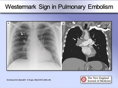
- CXR : hamptoms hump: wedgemark sign, westermark sign: pulmonary oligaemia in the affected segment


- CTPA : thrombi within pulmonary

https://epos.myesr.org/poster/esr/ecr2020/C-04154

- V/Q scan:
  • for those who have clot in the past, and have a previous episode before, or those who has acute kidney damage/ckd.
  • clear cxr
- ultrasound for lower limb: to rule out lower limb DVT due to long sitting/surgery...

- start DAPT

4. echo:
- look for RV dilation and hypokinesia. - acute rv failure--> increase afterload, rv cannot unload sufficiently causing rv dilated. impinge on LV cause LV to decrease output and supply to CA
- it increases o2 demand which CA could not meet, results in ischemia and necrosis

MASSIVE PE:
HYPOTENSION BP<50 for at least >15MINUTES, HR <40 - usually with evidence of MI and hyocardial dysfunction

Well's score for PE

low<2, mid 2-6, high >6
1. clinical signs of DVT -3
2. previous surgery / long traveeling>4hrs -1.5
3. previous DVT and PE- 1.5
4. HR>100 - 1.5
5. other diagnosis seem less likely -3
6.hemoptysis -1
7. cancer -1


Management (Mx):

anticoagulant
- without hypotension: LWMH, fonda/ DOACS
- unstable/ bleeding: parental anticoag 5-10d, switch to warfarin overlap 3-7d before discontinue heparin when INR >2 for >2d
- continue anticoag for 3-6months
thrombolytic therapy:
STK 25mu if pt high risk massive PE/ with cardiopulmonary arrest.
other mx: 
1. rivaroxaban 15mg BD for 21d then 20mg OD
2. dabigatran (expensive)

Sunday, February 14, 2021

Aortic Dissection

Intro:

-- Pathophysio
1. Risk Factors
2. Signs and symptoms
3. Blood Ix-initial preliminary ddx
4. ECG
5. Radiology 
6. Management
-- summary

Pathophysio

AD occurss when there is violation of the intimal layer of the aorta. This allows blood to dissect between the intimal and adventitial layers, causing the blood to flow into the media.

There are 3 possibilities as to how the blood enters the media:


  1. Atherosclerotic ulcer leading to intimal tear
  2. Disruption of vasa vasorum causing intramural haematoma
  3. De novo intimal tear

Following dissection, blood flow into the media may cause:


  • extension up or down
  • rupture
  • vessel branch occlusion
  • aortic regurgitation
  • pericardial effusion / tamponade


Risk Factors

1. inherited diseases: Marfan's
2. Underlying comorbidities: Hypertension, Hyperlipidemia, artherosclerosis
3. Others: Infection, history of cardiovascular surgery, arteritis, aortic dilation, aneurysm, pregnancy, trauma, old age
4. Iatrogenic: recent cadiac catheterization


Symptoms

1. severe pain at chest/back/abdomen/leg, depends on where the origin of aneurysm

2. signs: heart failure and hypotension

- unequal BP in the arms 20mmHg (suggest the presence of artery-clogging plaque in the vessel in limb with lower pressure)

- cold pallor /absent peripheral pulses in limbs 

- stroke/paraplegia

- anuria

- Aortic regurgitation

- left pleural effusion

 Murmurs

AR- early diastolic murmur
delayed distal pulse


Blood Ix

  • FBC:leukocytosis
  • Cr elevation with renal artery involvement
  • Tropnonin elevated : if dissection causes myocardial ischaemia
  • D-dimer – if negative --> dissection is very unlikely, but not sufficient to rule out
  • Cross-match: just in case there are excessive blood loss, in need of transfusion
  • Various biomarkers being investigated (e.g. elastin fragments, d-dimer, smooth muscle myosin heavy-chain protein)- if indicated


ECG

Results can be
  • normal
  • inferior ST elevation (right coronary dissection) but can be any STEMI (0.1% of STEMIs are dissections)
  • pericarditis changes, electrical alternans (tamponade)

example:

https://www.medscape.com/viewarticle/775982_2

The ECG shows findings of STEMI with right ventricular involvement (ST depression in lead I and elevation in VI). This could be a complication of an acute proximal aortic dissection, which can extend retrogradely, catching the proximal right coronary artery (RCA), either by a dissecting hematoma compressing the RCA lumen or a transsection of the proximal RCA. In either situation, an acute inferior STEMI can result as a complication of a proximal aortic dissection.

    Radiology

    • Chest X-Ray
    • CT angiogram
    • Ultrasound

    Chest X-Ray

    ** to always compare to old X-ray
    ** Loss of the aortic knob/aortic-pulmonary window and the calcium sign.
    ** widened mediastinum
    ** sometimes there are hemothorax /pleural effusion / loss of costophrenic angle (due to rupture)
    **pericardial effusion/tamponade/cardiomegaly


                                                                CT Angiogram (gold standard)

    reference CTA: 

    http://www.svuhradiology.ie/case-study/thoracic-aortic-dissection-ct/

    POCUS

    abdominal aortic dissection ultrasound

    Differential:


    Types of Aortic Dissection

    • Is it Stanford A / B

    TYPE A: 

      • severe and sharp chest pain 
      • --> surgical intervention
    • Involves ascending aorta. Can extend distally ad infinitum. Surgery usually indicated.


    TYPE B: back pain 

      •     (usually with malperfusion, ongoing progression, inability to control BP, and perforation.)
      • --> medical intervention
    • Involves descending aorta, aorta beyond left subclavian artery. Often managed medically with BP control.


    Management:

    Step 1:

    • O2
    • wide bore IV access (Swan sheath)
    • invasive monitoring
    • warn blood bank (x-match 6U + need for other products: FFP, PLT concentration, cryo) - depends on situation
    • correct coagulopathy

    Step 2:

    • control Pain
    Step 3:

    • control HR and BP (aim for P 60-80 and BP 100-120 SBP) target BP <120/80
      • Nitroprusside 0.25-0.5 mcg/kg/min then titrate or Nicardipine 5mg/hr
      • IV propanolol 0.5-1mg q5min till HR 60-80
      • Warning: Beware of pseudohypotension! of the bilateral BPs use the higher BP reading. if hypotensive, might lead to tamponade. resus with IV fluid and stop all anti-hpertensive agents

    • HR control:
      • IV beta blocker (propranolol, esmolol or labetalol) combined with vasodilators (e.g. GTN, labetalol, SNP)
      • start b-blocker first to avoid increased aortic wall stress from reflex tachycardia
    Step 4: 

    • call cardiothoracic surgeon if indicated


    Sample case for practice


    other cases: 

    https://www.ncbi.nlm.nih.gov/pmc/articles/PMC3856285/


    Reference:  

    1. type B AD

    2. https://emergencymedicinecases.com/aortic-dissection-em-cases-course/

    3. https://twitter.com/ManualOMedicine/status/1266758102667460609/photo/1