Saturday, June 26, 2021

OBGYN books for Houseman (HO)

Books recommended 
while having placement 
in Obstetric & Gynaecology 
as a (HO/houseman) 
in Malaysia

1. Clinical protocols in OBGYN for Malaysian
2. CPG Malaysia
3. HUSM labour suite - by Prof Adibah
4. Ampang notes O&G guideline
5. Quick management for gynae - by Dr Lee Say Fatt
6. Medicorp
7. Others


Protocols for OBGYN (Msia)
I find that it is a good guide for me when i am working OBGYN Houseman. A good book for those who practice as a doctor for hospitals in malaysia

Friday, June 25, 2021

EPR June 2021 OSCE

 Great thanks to those who contributed to the list, and hope you guys will be able to pass the exam with flying colours!

Good luck


EPR June 2021 (IMU)

1. IUGR/SGA ddx

  • - Small gestational age. 39th GW (counsellig) 

2. Nephrotic syndrome: 

  • Facial puffiness (hx taking, data intepretation, tx 

3. Megaloblastic Anemia

  • -PBF: hypersegmented neutrophil.Vit B12 deficiency

4. Suturing 

  • 3 simple interrupted suture)

5. ECG placements. 

  • Intepret Ecg ventricular tachycardia. how to use cardioversion?

6. Supraspinatus tendonitis

  • Shoulder physical examination. 
  • Xray-  Supraspinatus tendonitis,tx

7. compartment syndrome,

  • - Knee (full leg cast)  tx

8. Cataract

  • - Blur vision( hx taking) visual  acuity. Pupillary exam

9. PE neuro

  • Focal seizure (adult) hx taking, dx, two drugs

10. Pott's disease

  • Radiology (pulmonary tb, ddx) 

11. Alcohol abuse (hx taking) tx

12. Breaking Bad news

  • Patients family requesting not to disclose patient's terminal condition. Hepatocellular carcinoma

13. Hyperemesis gravidarum

14. ABCDE + ETT displacement

  • Correct asymmetrical lung intubation

15. Male urinary catheterisation

16. Neonatal jaundice, 

  • test for further Ix, graph chart for tx. ABO hemolysis? Physiological jaundice?

Postpartum haemorrhage


Sunday, June 6, 2021

Hypertension in Pregnancy

 Hypertension in Pregnancy

Differences

  • Chronic HPT: <20wks dx or beyond 6wks postpartum
  • Gestational HPT/ pregnancy induced hypertension (PIH without proteinuria) / Preeclampsia(PIH with proteinuria) / Eclampsia (PIH with convulsion): > 20wks dx
  • Chronic HPT with superimposed preeclampsia : PE in women who have pre existing hypertension. Include: worsen HPT, proteinuria, non-dependant oedema.


Pathophysio of preeclampsia

Found this video on youtube which is quite useful in helping to understand about Preeclampsia. Enjoy!

complications

Need for delivery when

- end organ damage
- inability to control BP
- fetal well being

Postpartum monitoring

- BP monitoring
- Ur protein and output checked
- check signs and Sx of preeclampsia --> postpartum eclampsia
- medication titrated according to BP
- counsel regarding subsequent pregnancies
- for contraceptions and proper spacing
- long term follow up at KK to watch out for chronic HPT. 

Management

1. Mild pregnancy induced hypertension:
  • diastolic BP 90-100, no proteinuria
  • Mx: 
    • rest at home with daily BP and urine check by community midwife
    • once or twice weekly Day Care Unit attendance for BP and CTG check
    • delivery by term or sooner
2. Moderate PIH     
  • diastolic BP 100-110 or less if complicated by proteinuria
  • Mx:
    • tx in hospital, daily ur albumin, 4hrly BP
    • check fluid balance, amount of oedema
    • weekly weight
    • Daily CTG
    • Meds: labetalol 100mg TDS max 300mg 6hrly , T. methyldopa 250mg QID to max 3g/d
    • deliver 38wks or sooner
3. Severe PIH
  • almost like severe PE Mx
    • proteinuria>3g/L
    • BP>160 systolic
    • BP>110 diastolic with no BP > than 160/110 with no proteinuria
    • oliguria (<400ml/24hrs)
    • presence of Impending Eclampsia
  • Mx:
    • Tx in hospital
    • Observation and meds like PIH
    • check for
      • reflex, clonus
      • opthal
      • LFT, PLT
      • quantitative proteinuria daily
    • Delivery by 36 wks or sooner
  • Aim to reduce to diastolic 90-100mmHg 

Key Points:

1st trimester :  1-12
2nd trimester: 13-26
3rd trimester: 27-EDD(40)

Proteinuria: 1+    0.3g/L,  
                      2+    1.0g/L, 
                      3+    3.0g/L, 
                      4+    >20g/L

Saturday, June 5, 2021

shoulder dystocia

 Shoulder Dystocia (SD)

1. Definition

2. Risk factors

3. HELPERR

4. BE CALM

5. Complications

6. Prevention

** HELPER, BE CALM

Definition: 

- unanticipated obstetric emergency which requires early recognition and rapid intervention

- refers to a situation where after delivery of the head, the anterior shoulder of the fetus becomes impacted on the maternal pubic symphysis, or (less commonly) the posterior shoulder becomes impacted on the sacral promontory.

RIsk factors:

  • maternal
    • maternal obesity
    • small maternal stature
    • GDM
    • previous SD 
    • IOL
    • prolong stage 1 and 2 of labour
    • augmentation of delivery
  • fetal
    • macrosomic
    • instrumental delivery
* those underline is the most common risk factors for SD. 

When SD occurs

1st: call for HELPPER!
H: call for HELP!
E: episiotomy
L: Mc robert maneuver


Mc Robert's maneuver: 
hyperflex and abduction of hips, cause cephalad rotation of symphysis pubis
flex mothers leg to abdomen,
 to flatten sacral promontory, straighten lumbosacral angle, increase A-P diameter of pelvis, 
thus free the impacted shoulder

P: suprapubic pressure: 
- help reduce bisacromial diameter, dislodge impacted shoulder, facilate to oblique position to permit delivery with gentle traction


E: Enter hand to deliver : 
- posterior arm delivery: finger introduce to fetal axilla to bring shoulder down and deliver posterior arm across baby chest

- wood screw method

- reverse wood screw

R: Roll on four (GASKIN maneuver)

all fail: for Zavenalli maneuver: atttempt to push back into uterus for Caesarean


Adapt BE CALM

- Breathe and stop pushing
- Elevate legs
- Call for help
- Apply suprapubic pressure
- enLarge vaginal opening: episiotomy
- Manoeuvres

Complications: 

  • Fetal:
    • Brachial Palsy, Brachial plexus injury, 
    • fracture of clavicla and humerus
    • cerebral hypoxia- d/t delay of delivery (should deliver within 5minutes once baby head out)
    • Ischemic encephalopathy
    • Cerebral palsy
  • maternal:
    • vaginal and perineal laceration
    • 3rd and 4th degree tears
    • postpartum hemorrhage
    • uterine rupture(rare)


Preventions:

- identify risk factors
- early IOL to prevent macrosomia
- Risk assessment: documentation of factors
- Early detection
- Plan of action: caesarean for babies > 4kg (in mothers with GDM), >5kg in mothers without GDM

References:

Friday, June 4, 2021

obstetric emergencies

Obstetric emergencies

1. Cord prolapse
2. Uterine rupture
3. PPH


Cord prolapse

Definition: 

1. overt umbilical cord presentation: umbilical cord lies infront of the presenting part and the membranes are intact. 

2. Overt umbilical cord prolapse: when the umbilical cord lies in front of the presenting part and the membranes have ruptured. 

3. occult umbilical cord presentation/ prolapse: the umbilical cord lies trapped beside the presenting part rather than below it

Risk Factors:

1. non- iatrogenic : fetal abnormal lie/malpresentation/breech, polyhydramnios, multiple pregnancy, prematurity, IUGR/SGA, high presenting part. 

2. iatrogenic: Amniotomy, placement of cervical ripening balloon catheter, vaginal manipulation of fetus with ruptured membranes.

3. External cephalic version. 

- complication: birth asphyxia (d/t cord compression or vasospasm)

Management:

- call for HELP! arrange OT!

- continue CTG

- relieve cord compression 

  •  using position: knee to chest position/ Tredelenburd position/ cephalad gravitation
  • help decrease blood flow, avoid fetal asphyxia and acidosis
  • cord vasospasm -which caused by exposing to surrounding
  • no oxytocin, only tocolytic agent: terbutaline 0.25mcg
  • ***do not push back the cord 
  • *** no amniotomy


- if you are doing VE: gentry push fetal head upwards, away from maternal pelvis (to relieve cord compression). use suprapubic pressure to keep fetus away from pelvis. 

- insert 500ml warm water in to urinig 

***1. discontinue oxytocin

        - administer tocolytic agents(s/c terbutaline 0.25mcg stat) if there are fetal brady

        - minimize excessive handling of the umbilical cord

        - if cord is outside, gently wrap the exposed cord with warm gauze. 

        2. never replace cord into uterus (causes vasopasm and fetal hypoxia)

        3. DONT remove examining fingers. 
        4. doppler ultrasound: to detect occult cord presentation

- deliver fetus ASoon/safeAP and 

- instrumental delivery if favourable

- breech extraction if favourable

- deflate bladder before peritoneal entry of C-section

Uterine rupture:

definition: separation of old uterine incision involving the entire thickness of uterine wall, with rupture of fetal membrane
- resulting in communication between uterus and peritoneal cavities

Uterine Dehiscence

- myometrial separation at site of uterine scar from previous surgery and uterine serosa remains intact. 

Causes:

1. spontaneous rupture, 
2. scar rupture, 
    - prior uterine surgery: C-section, myomectomy, 
    - D&C, hysteroscopy, forcep delivery, resection of uterine septum
3. traumatic rupture: 
    - uterine hyperstimulation (oxytocin- IOL)
    - obstructer labour: macrosomic baby, CPD
    - intrauterine manipulation (internal version, manual removal of adherent placenta)

Types:

1. completely rupture: extend through myometrium and serosal peritoneum

2. incomplete rupture: overlying peritoneum still intact, includes scar dehiscence

Symptoms:

  • PV bleeding 
  • suprapubic pain and tenderness
  • shock
  • undetectable fetal heart beat
    • CTG sudden cariable/late deceleration before onset of fetal bradycardia
  • easily palpable fetal body parts
  • loss of station
  • cessation of uterine contraction
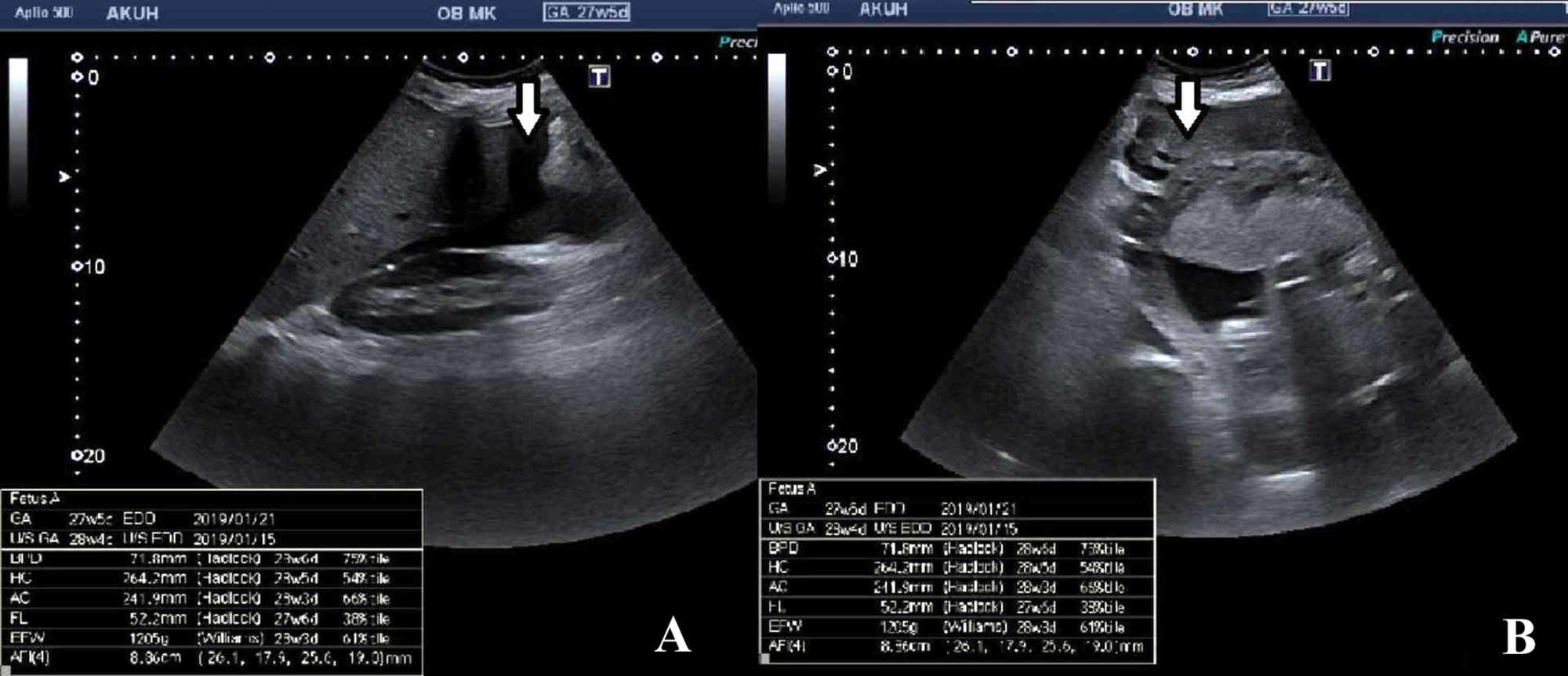
Diagnosis via

1. ultrasonography: 
  • protrude amniotic sac
  • hematome
  • endometrial/myometrial defect
  • intraperitoneal fetal part
  • hemoperitoneum/ free fluid

Management

  1. Intensive resus
  2. emergency laparotomy
    1. hysterectomy unless there are reasons to preserve uterus
    2. rupture repair
  3. Broad spectrum antibiotic
    1. cephalosporin
    2. flagyl (metronidazole)
  4. Adequate post operative care

POST PARTUM HEMORRHAGE

DEFINITION:

- Blood loss of more than 500ml following vaginal delivery or > 1L after Caesarean section. 
- Primary PPH: loss blood within 24hrs post-partum
- Secondary PPH: loss blood after 24hrs post-partum, within 12 weeks postpartum

Causes: 4T (tone, trauma, tissue, thrombin)

Mx: 

  • bleeding >1.5L --> can be seen in drop of BP
  • tachycardia >100bpm
  • 1st
    • active resus
    • uterine massage
    • IM symptometrine
    • 1ampule pitocin (max: 80u)
    • IM hemabate
    • then IVI 40u pitocin maintanence
*if uterine contracts after uteretonic drugs, continue with IVI of 40U pitocin in 500ml NS --> usually causes are uterine atony. 
*carboprost not given in bronchial asthma
* Hartmann 1L stat and check response (on acidosis, more lactate needed)
    - Hartmann's solution:  is a clear solution of sodium chloride, potassium chloride, 
                                      calcium chloride dihydrate and sodium lactate 60% in water.


Dose

Oxytocin

Ergometrine

15-Methylprostaglandin F2

Dose and route

IV : Infuse 20 IU in 1L

IV fluids at 60 drops/min

IM or IV (Slowly) : 0.2mg

IM: 0.25mg

Continuing dose

IV : Infuse 20IU in 1L

IV fluids at 40 drops.min

Repeat 0.2mg IM after 15 min

If required, give 0.2mg IM or IV (slowly every 4 hours)

0.25mg every 15 minutes

Maximum dose

Not more than 3L of IV fluids containing oxytocin

5 doses (Total 1.0mg)

8 doses (Total 2mg)

Precautions/ Contraindications

Do not give as an IV bolus

Avoid in pre-eclampsia, hypertension, heart disease

Bronchospasm (CI in Broncial  Asthma)


Tuesday, June 1, 2021

COPD

COPD

- pathophysio

a preventable and treatable respiratory disorder largely caused by smoking, is characterised by progressive, partially reversible airflow obstruction and lung hyperinflation with significant extrapulmonary (systemic) manifestations1 (Level II-2) and comorbid conditions2 (Level II-3) all of which may contribute to the severity of the disease in individual patients.


due to a mixture of small airway disease (obstructive bronchiolitis) and lung parenchymal destruction (emphysema), the relative contributions of which vary from individual to individual. Airflow limitation, associated with an abnormal inflammatory reaction of the lung to noxious particles or gases, the most common of which worldwide is cigarette smoke, is usually progressive, especially if exposure to the noxious agents persists. 

diagnosis and severity assessment of COPD, a post-bronchodilator FEV1 /FVC ratio of < 0.70 and post-bronchodilator FEV1 measurement, respectively are recommended.


The pathological changes in COPD, 

which include chronic inflammation and structural changes resulting from repeated injury and repair - -

- due to inhaled cigarette smoke and other noxious particles, 

- are found in the proximal airways, peripheral airways, lung parenchyma, and pulmonary vasculature.

The chronic inflammation in COPD is 

- characterised by 

  • an increase in the numbers of neutrophils (in the airway lumen), 
  • macrophages (in the airway lumen, airway wall, and parenchyma), and 
  • CD8+ lymphocytes (in the airway wall and parenchyma).
 The cells and mediators involved in the inflammatory processes in COPD and in asthma are different 

  • differences in physiological changes, symptoms and response to treatment 
  •  These pathological changes lead to 
    • mucus hypersecretion, 
    • expiratory airflow limitation 
    • with dynamic small airway collapse causing 
      • air trapping and lung hyperinflation, 
      • gas exchange abnormalities, and 
      • progressive pulmonary hypertension t
      • hat may lead to cor pulmonale.
    •  There is further amplification of the inflammatory response in the airways during exacerbations, 
      • which may be triggered by bacterial or viral infections or 
      • by environmental pollutants. 
  • In general, the inflammatory and structural changes in the airways increase with disease severity and persist on smoking cessation.


PE:

Physical signs of airflow limitation and air trapping (barrel chest, loss of cardiac and liver dullness, prolonged expiration, reduced breath sounds)

Ix


Signs symptoms of exacerbation

- >2 cardinal symptoms
: increased dypsnea/sputum volume/sputum prevalence

if 2/3--> moderate to severe
risk factors: age>65, fev1<50,>2exacerbation/year, with u/l IHD/ HF

noctural cough : why?

Patients with nocturnal asthma symptoms may have greater nighttime activation of inflammatory cells and mediators, lower levels of epinephrine and increased vagal tone


We can also use MMRC dyspnea scale and COPD staging to further improve our Management afterwards.

ddx: 

- bronchial asthma

- CHF

-pulmonary edoema,

pneumonia

-bronchiectasis

-pulmonary vascular disease


Outpatient management

- bronchodilators: beta agonist(salbutamol 2.505mg)/ipratropium bromide

- corticosteroids (fev1<50 30mg per day for 1week)


Types of meds

  • Acute exacerbation: doxycycline 100mg/ amoxycillin 500mg QID
  • CAP: ampicillin_ azithromycin
  • severe: 
    • wihtout beta lactamase inhibitor (piperacillin-tazobactam) /cefepime
  • Gram positive: 
    • staphylo, strepto, pneumococci, tb: cloxacillin, penicillin g,
  • gram negative: 
    • e coli, pseudomonas aeruginosa: aminoglycosides (gentamycin), cephalosporin(ceftriaxone), fluoroquinolones (ciprofloxacin)
  • tonsilitis: amoxycillin
  • meningitis: ceftriaxon + ampicillin