Saturday, February 27, 2021

common diagnostic criteria for medical

Criteria mentioned

if you cant remember can search https://www.mdcalc.com/ for auto calculation as well.

commonly used criteria in ward
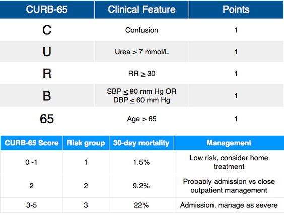

 Pneumonia - CURB 65

Confusion, urea>7, RR>30, BP<90/60, >65years old


Stroke risk - CHADS VASC 

- chronic heart failure-1, HTN-1,>75 years old-2, age 65-74 - 1, DMT2-1, stroke-2, vascular disease-1, female-1
-0: no antocoagulation needed, 1 : either no or with anti coag therapy, >2: oral anticoag theraphy recommended


Risk of bleed - HAS BLED

- HTN, Abnormal renal and liver fx(1 point each), stroke, bleeding, labile INR, Elderly >65, drugs or alcohol

- 0-2 low risk bleeding, >3 high risk bleeding, HTN : SBP>160


Peritoneal fluid - SAAG (serum to ascites albumin gradient)

serum albumin - albumin in ascitic fluid

- more useful than the protein based exudate/transudate concept.
>1.1 transudate/ indicates portal HTN (budd chiari, cirrhosis), <1.1 is exudate: look for inflammatory/neoplastic causes


Pleural fluid - Light's criteria

- to distinguish transudate or exudates

LDH/pleural fluid LDH, pleural fluid protein/ serum protein, pleural fluid/ serum LDH


Myocardial infarction - TIMI / KILIP

- thrombolysis in MI

TIMI: >65yo, >3risk factor of CAD, known CAD(stenosis>50%), ASA used past 7d

presented with recent chest pain<24hours, increase cardiac marker, ST deviation>0.5mm
- 0-2 low risk, 3-5 intermediate risk, 6-7 high risk


KILLIP: I: no congestion sign, II: with S3 basal rales, increase JVPIII: acute pulmonary edema, IV: with cardiogenic shock (bp<90/60, oliguria, cyanosis, impaired mental status)


NYHA: I: no symptoms and no limitations , II: mild symptoms and slight limitation III: significant limitation , comfortable at rest IV: severe limitation, Sx even at rest


PEmbolism Wells score

clinical signs of dvt, no alternate dx, hr>100, immobilise>3d, previous dvt/pe, malignancy, hemoptysis - 7 items


DVT well score: <2 DVT unlikely
- active cancer, paralysis, recent bedridden>3mth, localised tenderness, leg swollen, calf swelling, pitting edema on symptomatic leg, collateral superficial vein, previous dvt, alternate dx is at lease as likely as dvt (-2)


Padua risk assessment:


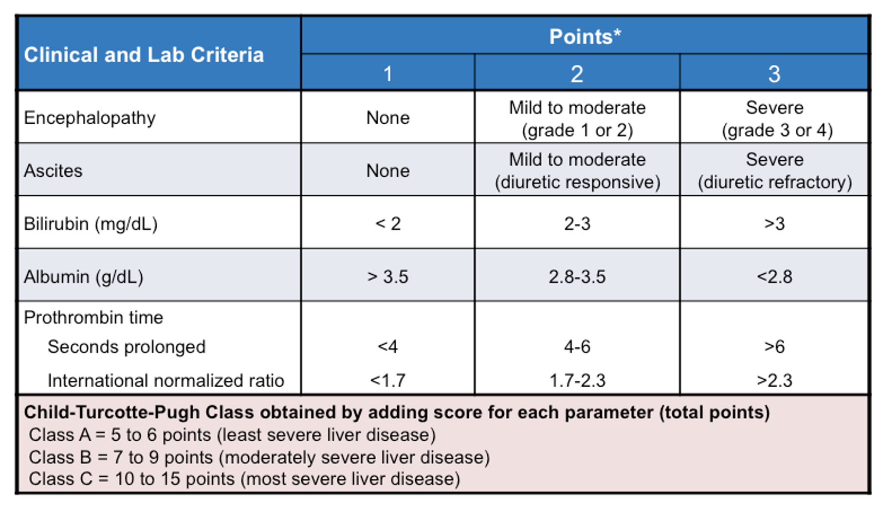
Liver cirrhosis - Child Pugh score

encephalopathy, INR>2/<1.9, Alb <2.8/>4, TB: >3.5/<1.8

10-15 child pugh C, 6-9 child pugh B, 1-5 child pugh A


GOLD ( COPD staging)

I-early, II-moderate, III-severe, IV-very severe


MMRC ( dyspnea severity)

0-4


Duke's criteria ( diagnose IE)

- 2major and 5 minor

- tp dx: at least 1major 1minor/ 5minor

- 2major: positive blood culture, endocardial involvement: echo positive for IE

-5 minor: fever>38, vascular phenomena, history of IVDU, immunologic phenomena, microbiological evidence

- 1 that rule them all is when Coxiella burnetii or antiphase I IgG antibody titer>1:800


Thyroid storm - Burch Wartofsky point scale

BWPS:>45 - definitive of thyroid storm, 25-44 clinical judgement needed


Mx: PTU 500-1000mg loading, then 250mg 4-6hourly - if cant give MMI 60-80mg/d

- high dose of IV HCT 100mg, 6hrly dexamethasone 2mg, 6hrly (to inhibit both thyroid hormone synthesis and peripheral conversion of T4 to T3)


Obstructive sleep apnea - STOP-BANG
- snoring, tiredness, observed apnea, high bp, bmi, age>50, neck circumference, male


ICH (mortality rate in ICB)

Intracerebral hemorrhage - GCS, age>80, location, ICH volume>30, intraventricular blood


NIHSS (assessment of stroke severity)
national institute of health stroke scale score: 11 item

LOC, gaze, visual, facial palsy, motor arm /leg, sensory, language, dysarthria, extinction n inattention


Framingham Risk Score (risk of MI)
- Age, HDL, FLP, BP, DM, smoker,


qSOFA (quick septic shock assessment)

- q sequential organ failure assessment


ABCD2 score ( stroke risk after TIA) 


EGSYS (identify cardiac syncope)

- evaluation of guidelines in syncope study

EGSYS

9 KPI for stroke patient 

9 KPI for stroke

Others:

1.Oxfordshire classification for stroke
2. CCS score ( severity of exertional angina)
3. Mayo DAI score ( severity of ulcerative colitis)
4. SLICC criteria ( diagnosis of SLE)


5. MELD score ( liver transplant assessment)
6. APRI score ( likelihood of9 fibrosis and cirrhosis in patients with Hepatitis C)
7. Lee index ( risk of perioperative cardiac events)
8. Epworth sleepiness scale ( to diagnose OSA) 
9. Mehran risk score (prediction of CIN)


10. Rheumatic fever (JONES criteria):

throat cultures: GABHS or elevated anti-streptolysin O titers + 2 major / 1major+2minor


Friday, February 26, 2021

Infective Endocarditis

Infective endocarditis

- common in pt with abnormal valves : rheumatic fever, valve surgery/replacement/ congenital abnormalities
- common bacteria: staphylococcus aureus, strepto, psudomonas aeruginosa, bartonella


Sample question:

A patient 40 years old male got admitted to the hospital, with fever for 7 days, will chills and rigor
Bp 120/80, pr 100bpm , T 38.5
Patient GCS 11, blur and unable to cooperate properly 
On physical examination with a pansystolic murmur heard on the left sternal border 
Presence of nodules on both arms

What is your diagnosis?

Ddx: Infective endocarditis

Peripheral signs: 
1. Nodes of the hand should prompt for more history: for example any needle injection history or not prior to this or it is just nodules. To differentiate between janeway lesion and osler nodes. 
- If they are IVDU highly suspected IE 
- generalised petechial as well due to bacterial peritonitis
- splinter hemorrhage in proximal nail plate
- osler nodes: cause by the infection in blood stream a type of neutrophilic vasculitis
- janeway lesion: bleeding in skin- staph aureus, microabscess from necrosis and inflammatory infiltrate--> on palms, soles, thenear, hypothenar and plantar surface of toes.

Central signs:
- hematuria: glomerulonephritis
- septic embolisation: lung embolisation
- Roth spots: red spot in the eye

Cardiac symptoms
2. Murmur heard at left sternal border
- tricuspid border is related to the tricuspid regurgitation- common on those heart valves with vegetative growth
- tricuspid valve endocarditis usually in drug abusers. 
    - damage by persistent bombardment of the endothelial surface with particulate matter from injected material 
- TR is accentuated with deep inspiration
- death usually from pulmonary regurgitation and RDS
or
- HF
- bradycardia

3. Fever most likely cause by the infection

Neurological symptoms
4. Drop of GCS 
- delirium - in meningitis/ encephalopathy
- confusion, drowsiness, reduced consciousness, vomiting, seizure - intracranial bleeding
- hemiparesis/aphasia: ischemic stroke

Any criteria to help with diagnosis of IE?

- Definite IE:
pathological criteria: 
    • microorganism demonstrated in culture/ histological examination of vegetation
    • pathological lesion: confirm by histological examination with active endocarditis
clinical criteria: 2major/1major and 3minor/ 5 minor
- Possible IE: 1 major and 1 minor / 3minor
- Rejected IE: firm alternate diagnosis/ resolution of symptoms <4days antibiotic/ does not meet criteria above

- DUKEs criteria: 
either 1 major 1 minor or 1 major 3 minor or 5 minor

Major
Positive blood cultures for IE
Evidence of endocardial involvement 
Minor
Predisposition: IVDU
Fever>38
Vascular phenomena: janeway lesion
Immunologic phenomena: glomerulonephritis
Microbiologic phenomena: positive blood culture that doesnt meet major criteria


Blood Ix to be sent 

- FBC: anemia, platelet count (thrombocytopenia)
- RFT: any acute kidney injury (a complication of IE)
- Electrolytes; to check if there are any depletion that causes the gcs drop
- LFT: any liver disease: hepatits 
- Infective screening
- Urine C+S: any hematuria or UTI
- CRP, ESR: infection
- coag: if pt on anticoagulation

- Blood C+S : 

1. to be taken 3 times from different sites before antibiotics/ treatment

- if remain negative for 5 days: consider negative
2. 3-4 days after commencement of treatment to document the eradication


Echocardiogram:

- irregular echo mass attached to valve
- if its tricuspid will relate to RV dilation, paradoxical septal motion and dilated IVC


ECG: what to look out for?

- Any changes for AV block or conduction abnormalities suggesting intracardiac extension of infection

CXR: Note multiple cavitating lung nodules due to septic pulmonary emboli.

- infiltration, cavitation, pleural effusion and empyema are common in S. aureus infection


Treatment: 

- Medical

- strepto: common among the young with  preexisting heart/ valve disease
- staphylo : common among old with healthcare related IE

while awaiting for investigation
- should give medical therapy first:
1. antibiotics with good coverage of staph aureus, streptococcus and pseudomonas
-6-8 weeks of parental therapy required. 


MONITOR treatment
- vital signs, regular examination
- daily ECG
- repeat echo once complete antibiotic (abx)
- review anticoagulant used

- surgical intervention

indications: 
1. persistent sepsis 
2. congestive heart failure
3. presented with cardiogenic shock
4. vegetation on echo >10mm in diameter


types of surgery: valve sparing debridement , valvulectomy with prosthetic replacement


Common complications:

1. Heart Failure: valve dysfx
- Diuretics (e.g. furosemide). 
  •  indicated for patients with symptoms of acute pulmonary oedema or fluid overload (raised jugular venous pressure; JVP and ankle oedema). 
  • Angiotensin converting enzyme (ACE) inhibitors. 
  • Beta-blockers: 
    • should also not be used in aortic regurgitation as this will increase diastolic time and regurgitation volume.

2. Persistent infection: causes AV block
3. Systemic embolism: occur in left sided IR within the first 2 weeks of therapy
- common sites are brain and spleen
4. neurological complications:
- early in course of IE (first 2 weeks)
- can be ischemic/hemorrhagic stroke/ aneurysm
    - to w/hold anticoagulation base on severity of complication

Prophylaxis

1. Antimicrobial prophylaxis not encouraged for cardiac patients
2. to consult with dentist prior invasive dental/medical procedure regarding the risk from IE
3. maintain good oral and skin hygiene

Reference:

Thursday, February 25, 2021

hypertensive meds

BP AIM:

BP for all hypertension pt: <140/90mmHg

BP for diabetics with nephropathy/ CKD/ IHD: < 130/80mmHg 

Hypertensive emergencies:

1. Aortic dissection: lower to<120/80, HR<60 in 1 hour

2. Hypertensive emergency: to lower 25% in first hour, and lower to 160/100 in 2nd to 6th hour, and normalise within 24-48 hours

3. Hypertensive urgency: to rest 30 minutes and repeat up to 2 hours, if doesnt decrease,  then lower BP 10-25% in 24hours not lower than 160/100mmHg. (captopril/nifedipine/labetalol)


Reference:

https://www.moh.gov.my/moh/resources/penerbitan/CPG/MSH%20Hypertension%20CPG%202018%20V3.8%20FA.pdf

Register for Covid 19-vaccination in Malaysia

Ever since the outbreak of covid-19, the world is never the same as before anymore. 
we are not allow to cross borders, no travelling, no mass gatherings and some could not even visit their family members!

Hopefully the situation of covid outbreak in malaysia could improve with the recent vaccination program kickstarted by the government yesterday. Our prime minister got his first shot of vaccine and more will get vaccinated as well.

I got curious and search up some articles regarding the pfizer vaccine and  here is some info you can read up regarding 

1. the pfizer vaccine. 
2. steps to register for vaccination in malaysia.
3. other information regarding the vaccine.


Get to know them better before getting vaccinated.

How vaccine can help contain diseases?
a picture is worth a thousand words

General info regarding the Pfizer vaccine

Vaccine type: mRNA , and does not contain eggs, perservatives or latex
it is given in 2 shots, 21 days apart
location: in the muscle of the upper arm

Consult the doctors if
- you had mild to severe allergic reaction to any ingredient in the covid 19 vaccine*
    - can develop either immediately or within 4hours of getting vaccinated
    - symptoms: hives, swelling, wheezing, shortness of breath
    - not allow to get the second dose of vaccine

Common side effects in a day or two after vaccine:
- might feel pain on the arm that you got injected
- can develop mild swelling or redness on the injection site
- some can have chills, tiredness or headache after getting vaccinated
- the side effects might affect your daily activites but should improve in a few days. 

Tips:
1. Keep calm and visit the doctor to talk about your pain or discomfort. Visit the doctor if the redness or tenderness worsens after 24hours.
2. Apply a clean, cool wet cloth over the vaccinated area that has discomfort
3. Drink plenty of water, make sure you are hydrated.


reference: 
https://www.cdc.gov/coronavirus/2019-ncov/vaccines/different-vaccines/Pfizer-BioNTech.html


Proses of vaccination through the mysejahtera App and website:


How to register: 

it is quite easy actually to get registered for vaccine

- in Malaysia, you can just register via My Sejahtera APP

- Steps are shown in diagram below

- check this website for more information as well: https://www.vaksincovid.gov.my/

- pdf download reference: vaccine program

- those without the app can register via

    1. the website

    2. all the hotline (coming soon)'

    3. at the public or private healthcare facility

    4. through assistance programmes for residents in rural and inland areas

reference: https://www.bernama.com/en/general/news.php?id=1932038


https://www.straitstimes.com/asia/se-asia/pm-muhyiddin-receives-first-covid-19-vaccine-as-malaysia-kicks-off-mass-inoculation

So what are you waiting for? 

Start registering via the app for the vaccine once you are ready!

Reminder:

Vaccinated ≠ stop wearing mask

However getting vaccinated doesn't mean you can stop wearing mask and be involve in mass gatherings to put ourselves and others at risk. This is because it is still unclear regarding which the vaccine can do to stop the infection and transmission. 

Be a responsible earth dweller and stay safe. 


Other Information regarding the vaccine:

the countries that have yet to start covid 19 vaccinations:

keep yourself updated here: 

Doctors discussion regarding vaccination:


References: 
3. regarding vaccinated ppl can skip covid quarantines
4. Vaccination for pregnant women
5. WHO also encourages for those at highest risk of serious disease or death to get vaccinated first against covid 19. They have been working on the ways to aim successful distribution of vaccine and treatments across all countries as well. link


Sunday, February 21, 2021

Diabetes and DKA

 Diabetes Mellitus type 2

that day we discussed with MO and he teach us something new, would like to share it to everyone here who is interested and also a reminder to myself. 

Question : how do we know when to give OHA or insulin in a patient.

Diagnosis criteria

- if symptomatic, one abnormal glucose is diagnostic
- if a asymtomatic, 2 abnormal glucose values required

  • symptoms of hyperglycemia
  • Fasting blood glucose: >7mmol/L
  • Random blood glucose : > 11.1mmol/L (at 2 occasions)
  • HbA1c: > 6.4 prediabetic

Targets

  • fasting BG: 4.4-6.1
  • non fasting 4.4-8
  • HbA1c <6.5
  • LDL<1.7, HDL<1.1, LDL<1.4(for pt with IHD) <2.6(for normal adult)
  • exercise: 150min/wk
  • BP: <130/80 (normal renal function), <125/75 (renal impaired)

Types of diabetic medication


New oral hypoglycemic agents (OHA):

- AGIs (acarbose): prevent carbohydrate absorption
- DPP4 inhibitor- sitagliptin: to excrete sugar from urine

reference: https://pubmed.ncbi.nlm.nih.gov/10989161/

Sunday, February 14, 2021

Aortic Dissection

Intro:

-- Pathophysio
1. Risk Factors
2. Signs and symptoms
3. Blood Ix-initial preliminary ddx
4. ECG
5. Radiology 
6. Management
-- summary

Pathophysio

AD occurss when there is violation of the intimal layer of the aorta. This allows blood to dissect between the intimal and adventitial layers, causing the blood to flow into the media.

There are 3 possibilities as to how the blood enters the media:


  1. Atherosclerotic ulcer leading to intimal tear
  2. Disruption of vasa vasorum causing intramural haematoma
  3. De novo intimal tear

Following dissection, blood flow into the media may cause:


  • extension up or down
  • rupture
  • vessel branch occlusion
  • aortic regurgitation
  • pericardial effusion / tamponade


Risk Factors

1. inherited diseases: Marfan's
2. Underlying comorbidities: Hypertension, Hyperlipidemia, artherosclerosis
3. Others: Infection, history of cardiovascular surgery, arteritis, aortic dilation, aneurysm, pregnancy, trauma, old age
4. Iatrogenic: recent cadiac catheterization


Symptoms

1. severe pain at chest/back/abdomen/leg, depends on where the origin of aneurysm

2. signs: heart failure and hypotension

- unequal BP in the arms 20mmHg (suggest the presence of artery-clogging plaque in the vessel in limb with lower pressure)

- cold pallor /absent peripheral pulses in limbs 

- stroke/paraplegia

- anuria

- Aortic regurgitation

- left pleural effusion

 Murmurs

AR- early diastolic murmur
delayed distal pulse


Blood Ix

  • FBC:leukocytosis
  • Cr elevation with renal artery involvement
  • Tropnonin elevated : if dissection causes myocardial ischaemia
  • D-dimer – if negative --> dissection is very unlikely, but not sufficient to rule out
  • Cross-match: just in case there are excessive blood loss, in need of transfusion
  • Various biomarkers being investigated (e.g. elastin fragments, d-dimer, smooth muscle myosin heavy-chain protein)- if indicated


ECG

Results can be
  • normal
  • inferior ST elevation (right coronary dissection) but can be any STEMI (0.1% of STEMIs are dissections)
  • pericarditis changes, electrical alternans (tamponade)

example:

https://www.medscape.com/viewarticle/775982_2

The ECG shows findings of STEMI with right ventricular involvement (ST depression in lead I and elevation in VI). This could be a complication of an acute proximal aortic dissection, which can extend retrogradely, catching the proximal right coronary artery (RCA), either by a dissecting hematoma compressing the RCA lumen or a transsection of the proximal RCA. In either situation, an acute inferior STEMI can result as a complication of a proximal aortic dissection.

    Radiology

    • Chest X-Ray
    • CT angiogram
    • Ultrasound

    Chest X-Ray

    ** to always compare to old X-ray
    ** Loss of the aortic knob/aortic-pulmonary window and the calcium sign.
    ** widened mediastinum
    ** sometimes there are hemothorax /pleural effusion / loss of costophrenic angle (due to rupture)
    **pericardial effusion/tamponade/cardiomegaly


                                                                CT Angiogram (gold standard)

    reference CTA: 

    http://www.svuhradiology.ie/case-study/thoracic-aortic-dissection-ct/

    POCUS

    abdominal aortic dissection ultrasound

    Differential:


    Types of Aortic Dissection

    • Is it Stanford A / B

    TYPE A: 

      • severe and sharp chest pain 
      • --> surgical intervention
    • Involves ascending aorta. Can extend distally ad infinitum. Surgery usually indicated.


    TYPE B: back pain 

      •     (usually with malperfusion, ongoing progression, inability to control BP, and perforation.)
      • --> medical intervention
    • Involves descending aorta, aorta beyond left subclavian artery. Often managed medically with BP control.


    Management:

    Step 1:

    • O2
    • wide bore IV access (Swan sheath)
    • invasive monitoring
    • warn blood bank (x-match 6U + need for other products: FFP, PLT concentration, cryo) - depends on situation
    • correct coagulopathy

    Step 2:

    • control Pain
    Step 3:

    • control HR and BP (aim for P 60-80 and BP 100-120 SBP) target BP <120/80
      • Nitroprusside 0.25-0.5 mcg/kg/min then titrate or Nicardipine 5mg/hr
      • IV propanolol 0.5-1mg q5min till HR 60-80
      • Warning: Beware of pseudohypotension! of the bilateral BPs use the higher BP reading. if hypotensive, might lead to tamponade. resus with IV fluid and stop all anti-hpertensive agents

    • HR control:
      • IV beta blocker (propranolol, esmolol or labetalol) combined with vasodilators (e.g. GTN, labetalol, SNP)
      • start b-blocker first to avoid increased aortic wall stress from reflex tachycardia
    Step 4: 

    • call cardiothoracic surgeon if indicated


    Sample case for practice


    other cases: 

    https://www.ncbi.nlm.nih.gov/pmc/articles/PMC3856285/


    Reference:  

    1. type B AD

    2. https://emergencymedicinecases.com/aortic-dissection-em-cases-course/

    3. https://twitter.com/ManualOMedicine/status/1266758102667460609/photo/1