Monday, November 15, 2021

HPB Q&A for HO

HPB = Hepatobiliary

Anatomy of HPB 


Common classifications used in HPB

1. Ranson criteria


2. Mirrizi classification


biliary colic -> acute cholecystitis -> acute cholangitis, pancreatitis, gb empyema and gangrenous gb

presentation of each spectrum

Causes of obstructive jaundice


painless vs painful jaundice

  • Painful obstructive jaundice is usually related to gallstones, while painless obstructive jaundice tends to be related to tumours.


Q1: If patient came with features of ascending cholangitis, what Investigation you would like to order

  • FBC - to monitor TWC, PLT. 
    • low platelet: sepsis induces/ severe sepsis that causes coagulopathy
  • BUSE - urea, creat monitoring
    • hyperbilirubinemia can cause hepatorenal syndrome and renal impairment
    • severe sepsis that causes organ failure : kidney failure
  • LFT: TB, kiver enzymes and albumin level
    • especially ALP and direct bilirubin that can help determin if there are any obstructive jaundice
  • ABG: any acidosis or resp failure
  • CXR first, as we can use it to rule out perforated viscus
    • unless obvious jaundice, so less likely perforated PUD

Q2: why we need ultrasound HBS for ascending cholangitis?

    • to rv CBD(common bile duct) size, and any dilatation
    • any stone present- if there's obstruction , might need urgent intervention to relieve obstruction
    • sometimes stone cant be seen as obscured by bowel gas. 
      • stone is usually in distal CBD - the narrowest part
      • and it is at the back of duodemun
      • usg wave cannot go through gas
    • To rule out liver abscess
      • as the presentation of liver abscess is the same: RHC pain, fever and jaundice

Q3: normal size of CBD?

  • 3-5mm
  •  >8mm considered dilated (usually we use >10mm or 1cm as dilated)

Q4: why do we need to rule out liver abscess urgently?

  • if huge liver abscess need to drain urgently. 
  • gold standard tx: percutaneous drainage and antibiotic
  • otherwise it will rupture and cause peritonitis --> severe sepsis --> mortality

Q5. if found out patient have ascending cholangitis secondary to choledocholithiasis, how would you manage?

  • resuscitate and stabilize the patient first with ABC
  • Airway
  • Breathing
  • Circulation
  • insert branula + blood for Ix and blood culture
  • fluid resus
  • antibiotics
  • analgesia

Q5a. what are the choice of antibiotics use for the pt above?

  • HBS common organism are
  • function of liver: it uses portal circulation, 
    • it collects blood from gut  - all the guy -> return back to liver
  • therefore in the GI tract, mainly organism gram negative and anaerobes
  • also called BROAD SPECTRUM of negative organisms
    • so we need at least 3rd generation cephalosporin 
    • eg: cefobid / cefoperazone
  • to cover anaerobes: metronidazole

Q6. after analgesics what can we do for patient with ascending cholangitis and in pain?

  • if Stable, we can try ERCP to remove the stone . if unable to do so we will put stent to drain the obstruction
    • ERCP needs sedation, and prone position. therefore difficult to maintain airway
  • If patient is unstable , the option is PTBD

Q7. when to do cholecystectomy?

  • once sepsis is resolved
  • stone not cleared through ERCP 

Q8. How do you tell which liver segment was it from a CT scan?

Anatomy of liver :
note that 2 is beside 4a, 3 below 2 and 4b is below 4a
upper segment at right side is segment 7,8
and lower right is segment 6 and 7


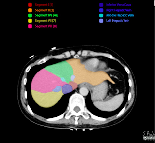
 First you can check the segments from identifying the hepatic veins. As shown in diagram above, left hepatic vein separates segment 2 and 4a, then the middle hepatic vein for segment 4a and 8, right hepatic vein for segment 8 and 7


Then as you scroll down the ct scan, you will see the portal vein (left branch). the portal vein separates the liver to the left(segment 2 and 4a) and right side (segment 7,8) . below the portal vein emerges segment5


When you can see the right branch of portal vein, it also indicates that on Left (segment 4b, and segment 3), on its right is segment 5 and segment 6


reference, more on website: 


Saturday, November 6, 2021

Electrolyte Imbalance QnA for HO

 House Officers must know in ward 

- Electrolyte imbalance


Q1: 50year old, 70 kg, male with persistent diarrhea and vomiting for 3days due to food poisoning. He is dehydrated. Vitals are stable, looks lethargic. Na: 128mmol/L

a. How to correct his hyponatremia

    - calculate first
  • Na deficit (mmol) : (desired Na level - serum Na) x bw x fraction
  • 0.6 x kg x (desired Na - serum Na)
    • desired Na :  135-145
    • Na deficit is 0.6 because extracellular comprises 60%. 
    • If its potassium (intracellular) therefore 0.4 cause it is 40%
  • 0.6 x 70 x (135-128) = 294mmol/L
    - to add with daily requirement
  • infusion = deficit + requirement
    • requirement: 1-2mmol/kg/d (we usually use the lower range when calculating)
  • infusion: 294 + 70 = 364 mmol/L
- then how do we deliver 364mmol/L to pt?
  • 1 pint NS = 77mmol/ L
  • 1L NS = 154mmol/L
  • 364 / 77 = 4.7 pints, 
  • therefore 5 pints of normal saline over 24 hours is needed.

b. the next day his repeated Na is normal: 136, 

how do you plan for fluid regime ? for maintenance?

  • normally a person need 30-40ml/kg/d (fluid requirement)
    • general normal weight for man: 70kg,  normal weight for woman : 60kg
  • if we take the middle value fluid requirement (35ml/kg/d), 
    • the man will need 2450ml (around 5 pint as well)
    • women is 2100ml, around 4pint. 
thats why usually we see usually 
  • 5 pint (2/3 pint NS + 2/3 pint D5%) given to man, 
  • 4 pint(2 pint NS + 2 pint D5%) given to women. 
** WHY 2 or 3 pint NS? for example a 70kg man we take 140mmol Na requirement, 1 pint NS is 77mmol, therefore the man only need 2 pint NS. but his fluid requirement is 5 pints a day, that's why we need to add another 3 pint D5 to complete his daily fluid requirement. 


Q2. what should you watch out during sodium correction?

  • Na correction should not exceed 10mmol/L/24 hours
    • thats why we use the lower normal when correcting sodium
  • even in severe hyponatremia, if Na only 120, your aim of correction is only up to 130, and not up to 135 in 24 hours. 
  • if there are hypernatremia (rare), mostly we try to find out the associated conditions/ related underlying issues. usually will be given diuretics to excrete excess sodium. 


Q3: 50year old, 70 kg, male with persistent diarrhea and vomiting for 3days due to food poisoning. looks lethargic, no ecg changes.  K: 3.2mmol/L

a. how to correct his potassium?

  • Deficit = (desired K - measured) x Body Weight (BW) x 0.4
    • (3.5-3.2) x 70 x 0.4 = 8.4mmol
    • 70 for maintanence , 
    • maintanence + deficit = infusion
      • 70+8.4 = 78.4
    • 78.4/13.3 = 5.8gm
      • we take normal daily requrement of K : 1mmol/kg/d, therefore we need to convert into gm  by dividing 13.3
      • 1gm = 13.3mmol
    • so we need to correct at least 5 gm first
      • can correct with 1g KCL in each pint NS (total 5gm)/24hours

b. what is the maximum/ safe dilution of K+ in 1 pint of NS?

  • dilution cannot exceed 40mmol/L
  • which also means cannot exceed 20mmol/pint = 20/13.3 = 1.5gm KCl
  • we cannot give more than 1.5gm KCl in a pint of NS

c. what is the safe titration of K+?

  • titration of potassium cannot exceed 10mmol / hour
  • if we need fast correction due to symptomatic, 
    • max is 1gm KCl in 100cc NS over 1 hour, not faster than that
  • If need 2gm infusion, it must be in 2 hours.
    • never bolus correction, must always use safe titration
    • 2gm KCl infusion in 200cc NS over 2 hours
  • OTHERWISE can cause CARDIAC ARREST! 

Q4: How do we know the patient has hyperkalemia?

  • Cut off point for hyper K: 
    • K+ > 5.5
    • symptomatic ECG changes: tall tented T waves, absence of P wave, broad QRS, PR prolong
    • it became dangerous when the ECG changes go towards heart block type of ECG with tall tented T wave --> severe hyperkalemia --> lead to arrythmia then asystole

Q5. what is the fx of each components in lytic coctail? 

  • lytic cocktail: treatment for hyperkalemia
  • Calcium gluconate - 10ml 10% calcium gluconate
    • for cardio protection
  • Insulin / actrapid 10u : 
    • drive K+ into cell together with glucose
  • Glucose/ D50 - 50ml : 
    • K+ transporter
  • we need to administer calcium gluconate follow by insulin then D50

Q5a. so why we cannot give insulin first instead of glucose?

  • Potassium cannot enter the cell by itself, thats why we need glucose first ,since it is the transporter it can allow the insulin to drive the K into the cell. 
  • GLucose is the transporter
    • so we need to load the excess potassium onto the glucose
  • Insulin cannot push the K directly, they need transport. therefore they need glucose first, then insulin will push both into the cells. 

Q5b. how many times can we give lytic cocktail?

  • every 6 hourly
  • if the potassium is still not corrected, Hemodialysis (HD) is needed

But we could not just correct potassium with just lytic coctail,  have to correct the cause of hyperK as well. 

Q5c. what are the common cause of HyperK in surgery?

  • Acute Kidney Injury (AKI)
  • severe dehydration, sepsis can cause AKI. 
  • If there are evidence of sepsis, we need to find the source and remove its source of infection
    • antibiotics
  • If there are acidosis, we need to find the cause and solve it from there as well. 

Q6. what are the common antibiotics used in Gastrointestinal - GI sepsis?

  • cefobid and flagyl
  • common organism in GI
    • gram negative : E.Coli, Klebsiella, Enterobacter
    • GI accomodates a broad spectrum of gram negative organisms
    • anaerobes
  • 1st generation of cephalosporin covers gram positive
    • exp: cephalexin
  • 2nd generation covers gram positive and a bit of gram negative (narrow spectrum)
    • cefuroxime
  • 3rd generation covers broad spectrum of gram negative
    • cefobid/ cefoperazone
  • Metronidazole: covers anaerobes
  • If patient has carbuncle, abscess or soft tissue infection / sepsis, the choice of antibiotic should be covering skin organism: 
    • empirical antibiotic
    • penicillin based: cloxacillin, etc. 
  • for Urology patient, usually have narrow spectrum of organism, 
    • common: E. Coli
    • can use cefuroxime as empirical antibiotic, unless they have sepsis, so may need to consider quinolones to cover for broader/ other pathogens

Q6a. so when do we change antibiotics? 

  • observe if antibiotic works, after 3-5 days
  • usually the patient will show clinical improving and total white cell count (TWC) will reduce
  • but if no changes after 3 days, we might need to consider changing to a higher efficacy antibiotic


Friday, November 5, 2021

Fluid and Resus common QnA for HO

Part 1: Fluid and its components

Q1. What are the common cause of fluid loss in surgery?

  • apparent loss: diarrhea, vomiting and high output stoma
  • 3rd space loss: 
    • loss of water, electrolyte and colloid particles into interstitial space
    • which could contribute to edema
    • Intestinal obstruction, pancreatitis and ascites
  • others: insensible fluid loss (hyperventilation/pyrexia), stress response

Q1a. How patient loss fluids from IO?

  • apparent loss: vomiting
  • 3rd space loss: 
    • increased secretions
      • bowel obstruction will cause bowel to secrete a lot of secretion to overcome the obstruction. 
    • mucosal edema, so fluid not absorbed
      • so there will be a lot of accumulation of fluid that leads to third space loss
      • fluid accumulation in bowel can reach up to 6L
      • that could lead to hypovolemic shock

Q1b. How patient loss fluid from pancreatitis?

  • systemic inflammation 
    • inflammation causing release of inflammatory cytokine and other pro-inflammatory mediators,
    • leading to capillary leakage
    • thus loss of circulatory albumin and fluids to interstitium
      • capillary leakage cause fluid shift to third space and then hypotension leads to hypovolemic shock.

Q2. What is the main difference between crystalloid and colloid?

  • molecular size
    • affects shifting of fluid where low molecular size - low tonicity


Q3. How does fluid moves in human body?

  • from low concentration to high concentration 
    • big molecules fluid (colloids) 
    • has ability to pull fluids from other compartments : oncotic pressure
  • opposite of oncotic pressure: hydrostatic pressure
** not through pressure gradient / osmosis

Q4. What is isotonic?

  • a solution concentration that is similar to plasma
  • the osmolarity of plasma is around 300 mosm/L
    • Normal saline: 308
    • Half saline: 154 (hypotonic)
    • 3% saline: 1026 (hypertonic)

Q5. What is the difference between Normal Saline (NS) and Hartmann(HM) solution?

  • Hartmann has additional potassium, lactate, calcium
  • the most "physiological" solution

Q5a. What is the function of lactate in HM

  • lactate will be metabolised by liver to HCO3, thus acting as buffer
    • especially in met acidosis

Q5b. Can we use HM as resuscitation fluid?

  • No
    • usually when patient needs resuscitation, already with multiple organ failure or impairment
    • so although Hartmann has lactate that could help as buffer, the liver is unable to convert lactate to bicarbonate, which could lead to accumulation
    • the accumulation of lactate will worsen the metabolic acidosis
    • and Pt in acidosis usually has hyperkalemia, Hartment contains K+ which would further worsen the situation


Q5b. So when do we use hartmann?

  • as maintanence, especially in those who need replace electrolyte loss (diarrhea and vomiting)

Q6. What is the function of Dextrose 5%?

  • provide hydration
  • the have glucose in solution not for calorie to avoid lysis and avoid hypotonic
    • calorie in D5: 170/L
  • it is just to render solution isotonic once infused in the circulation, once they reach liver will convert into free fluid
    • - provide free water that can pass through membrane pores, expanding both intracellular and extracellular spaces


Part 2: Fluid and resuscitations

Q7. 60year old, 70kg man presents with diarrhea and vomiting for 1 week. brought in with hypovolemic shock
Outline your management for this man

- ABC
  • assess airway
  • breathing
  • circulation
    • check the vital signs: unstable/ stable
- insert 2 large bore needles and give IV NS
- run fast 1 pint NS

Q7a : If patient doesn't respond to fluid resus? 

  • reaccess: if the volume is improved, but patient still hypotensive, he might have other component of shock
    • for example: septicaemic shock --> we might need to start inotrope for vasoconstriction
    • if cardiogenic shock / has underlying IHD --> get an ECG, and we might need to start with dopamine or dobutamine as the inotropic support. 
  • if volume is still low, 
    • can infuse COLLOID to hold the fluid in the circulation
    • colloid has oncotic pressure that will hold the fluid intravascularly, thus maintain the BP
  • So why we cant give colloid straight away for resus?
    • Colloid causes shifting of fluid out of the cell, worsen the hypoperfusion
    • in shock, circulation fails and tissue is hypoperfused, if we infuse hypertonic solution all fluid will move from the tissue into the intravascular system . 
    • Therefore load with volume first (crystalloid)
      • resume the circulation
      • let them reach the heart, brain and kidney
      • after that infuse colloid to hold the volume. 

Q8: Define shock

  • must have 2 components
    • circulatory failure: seen via vital signs
    • inadequate tissue perfusion : seen via low SPO2
      • sequelae of low perfusion
      • multiorgan failure

Q9: Why we cant have central line when patient is in shock?

  • it is about the catheter's caliber. the shorter the calibre, faster the infusion. 
    • if central line, it has long calibre and the rate of infusion is slower
    • insertion takes a lot of time
  • in shock we need large supply of fluid for the patient 
    • Poiseuille law

Q10: How do you know patient responded to your fluid resuscitation? what are your AIMS?

  • vital signs 
    • HR <90
    • BP >90/60 , MAP >60
    • SPO2 >95%
    • RR <20
    • u/o  >0.5ml/kg/hr