Sunday, January 7, 2024
EM Medications
Saturday, July 23, 2022
Paediatric as HO
Paediatric books to study in HO
In our hospital when I am in paediatric department posting, mainly using the paediatric protocol book. It is quite important as we use it when we have presentations in department.
Other than paed protocol, I found that the following other books and website is also useful.
Neonate Paediatric / Oncology paediatric:
Paediatric Protocol
MOH Paediatric Protocols for Malaysian Hospitals, 4th edition
General Paediatric:
Ampang guideline
https://myhow.files.wordpress.com/2013/11/hoguidepeds1.pdf
Physical examination:
MRCPH website
https://mrcpch.paediatrics.co.uk/
Medications
Things to prepare:
- Pens
- Measuring tape
- Mini calculator (either those pocket size or the ones that you can hang on your landyard)
- Scissors
- Durapore
Notes to bring along
- Fluid requirements
- Growth development
- Growth chart
- Normal readings of blood test
- Normal values for vital signs (RR, BP percentile)
- it is quite challenging and difficult to memorise the whole chart, easier to bring it around and refer when needed.
- mini toy/ cute things
- (to help kids to calm down before any procedures, eg: taking blood/ punctures)
- I have a mini duck with me most of the time, and i find it very useful in making the kids happy and cooperate. However later on I realise they just like anything that is in bright colour, not necessarily toys. Even a cute plaster could make them feel so happy.
Good Attitude
- kids are often affected by our mood/ emotions, so do make sure you are treating them with joy, patience and love.
Sunday, July 10, 2022
Abdominal examination for paeds
Abdominal examination:
General Examination
- General surroundings
- equipments : NG tube/ PEG tube
- nutrition: milk, food
- Growth chart
- Hand
- finger clubbing
- crt, pulse volume, warm/cold peripheries
- rashes?
- Face
- eyes: anemia? edema?
- mouth: gum bleed/ hydration
- cyanosis
- overall inspection
- Neck
- spider naevi
- inflammation/ edema on the neck
- lymph node
- Abdomen
- Inspection
- shape
- distension
- masses
- rashes/scar/striae/stoma
- Palpation
- pt should be in supine position, gently ask if it is comfortable to lie down if having abdominal distension
- superficial, deep palpation
- liver/ spleen/ kidney palpation/ abdominal mass
- Percussion
- shifting dullness/ fluid thrill
- Auscultation
- resonance/ dull
- Lower back
- sacral edema?
- Lower limbs
- pedal edema
- Nephrotic syndrome : ascites/ periorbital edema/ pedal edema/ sacral adema/ pleural effusion
Causes of hepatomegaly
1. Structural: Extrahepatic biliary atresia, choledochal cyst, intrahepatic biliary hypoplasia, congenital hepatic fibrosis, polycystic disease
2 . Storage/ metabolic:
– Carbohydrate- glycogen storage disease (Type 1,3,4,6), hereditary fructose intolerance, galactosaemia,, mucopolysaccaridoses-
– Mineral: Wilson’s disease, juvenile haemachromatoses
-Nutrition: Protein calorie malnutrition, TPN-Bile flow: progressive familial intrahepatic cholestaisis syndrome
-Protease: Alpha-1-antitripsin
-Electrolyte: CF
– Amino acid: Tyrosinaemia type 1, urea cycle disorder
-Lipid – Gaucher, Nieman-Pick diease, cholesteryl ester storage disease
3. Haematological: Thalassaemia, sickle cell disease, ALL, AML CML
4. Heart/ Vascular: Congestive heart failure, constrictive pericarditis, obstructive IVC, Budd-Chiari syndrome
5. Infection: Viral – rubella, CMV, coxsackie virus, echovirus, hepatitis ABCDE, EBV ; Bacteria – E.coli TI, TB, syphilis ; Parasite – Malaria, toxoplasmosis, Schistosomiasis
Monday, June 13, 2022
Cardiovascular examination for children
Some physical examination compilation of videos to help with revision
Cardiovascular examination
Introduce yourself and ask the kid politely for some information that could aid in your diagnosis later
- age, growth chart, development milestones
- history - any dyspnea/palpitations, exercise intolerance, dizziness/ syncope, family hx (Marfan, chromosomal, CHD in 1st degree relative)
- any vital signs?
- current complain
Next, we go to physical examinations
- general examination (cyanosis, dysmorphic features)
- peripheral pulses (finger clubbing, pulse volume, capillary refill - radial/carotid/femoral)
- check for any signs of anaemia, dehydration
cardiovascular examination:
- palpation - apex/ heave/ thrill
- auscultation - lying down, sitting up forward, hold breath
- timing (systolic/ diastolic)
- quality (soft/ harsh)
- intensity- grade II (innocent), grade III or higher (possible thrill)
- louder with exercise / anaemia / fever / position?
- any other sounds?
- check for edema - sacral/ leg
- hepatomegaly (common in heart failure children)
Friday, April 22, 2022
Common Questions about Intestinal obstruction (IO)
1. What are the cardinal symptoms of IO?
-, colicky abdominal pain, abdominal distention and vomiting, no BO, no flatus (obstipation/ absolute constipation)
- other history you want to establish
- chief complain
- to explore regarding the cardinal Sx
- duration of symptoms
- abdominal pain: to see the location so we can locate either small or large bowel.
- if its small bowel pain : felt in the upper abdomen and central (periumbilicus) while if large bowel pain is felt in lower abdomen
- these are all visceral pains that is why you referred to area of referred pain
- small bowel first
- proximal, smaller in diameter- which would cause distension early, higher level of obstruction
- small bowel is also active in peristalsis, so once they are obstructed they will cause intense colicky pain.
- onset :
- small bowel is active in absorption of nutrients in GI tract. It produces a lot of fluids. If it is obstructed there will be a lot of fluid accumulation so the vomiting will occur early.
- while in large bowel there is not much of fluid and it is distal, the vomiting occurs late
- content : we will be able to observe the content either from vomiting, or from an NG tube insertion. then we can see the colour content in the bag
- if its feculant material : lower small bowel
- greenish or bile content : high small bowel
Q2. causes of IO and how do you classify
- dynamic
- adynamic
- first we need to rule out mechanical obstructions first
- intraluminal : tumor
- impacted stool: rare, usually in pt with hirchsprung disease
- colon tumor: left sided tumor (Large bowel obstruction)
- small pellet stool, constitutional symptoms, blood in stool
- bowel habit
- PR bleed
- mucous discharge
- tenesmus
- family hx of colon Ca
- intramural,
- extramural : adhesion, hernia
- hernia usually affects the small bowel. so if the patient with cardinal symptoms mention above leading towards SBO, remember to confirm regarding their abdominal surgery and hernia.
Q3. Complications of IO
- perforation
- pain became severe and intolerable
- localised pain became generalised
- presence of high grade fever
- abdomen become more distended
Wednesday, March 16, 2022
Ortho HO must know
Clerking key points:
- age/ race/gender
- chief complain
- presenting history (how it happens-where they fall, how, why; pain-what kind of pain-location, intensity, relieving factors)
- other related signs and symptoms: LOC(loss of consciousness), headache, N&V(nausea and vomiting)
- treated by any other hospital?
- past medical history: underlying disease? DM/HPT? well controlled?
- Social hostory: ADL, who he is staying with?
- P/E- relevant and related: Observe, palpate, motor: Range of movement of limbs, sensitivity, neurological
- report any X-ray or CT scan done
Review pt in the morning.
- the header is S/B Dr. XXX or <AM review>
format:
- age/race/gender
- Post operation Day/ Day admission, what kind of surgery,
- pt condition: oral and toiletry, SOB, fever, cough/flu, pain(pain score),
- vital signs: BP, Pulse, T
- P/E: dressing, wound/condition
- Plan: continue previous meds, change dressing, physio referral, feeding, pain medication
Must know: (for ortho)
- how to read the spine X-ray/ any xray
- check alignment
- body: vertabral body, spinous process, antevertebral body, 4 lines of X-ray,
- any burst /compression fracture
- to determine via TLCIS if the person with thoracolumbar injury needs surgery or not.
- physical examination
- motor, sensory and neurological: any abnormalities, know what is it testing for
- differences between Upper Motor neuron and Lower Motor Neuron,
- Cauda Equina
- end of UMN is L2
COMMON TO DO LIST
- TO do ABG, use the blue needle and syringe to extrct the blood
- to do line, use pink needle, find the vein on pt hand, feel and put it in.
- to do slap, need a friend to help with releasing the water from the back slap(10 layers)
- to do skin traction: prepare the cover and weight(10% of pt body weight)
- to be prepared always for mo who is coming for pt.
Friday, February 11, 2022
fracture classifications
Common classifications used during orthopedic posting.
In a glance:
Gustilo classification: open fracture
Gartland classification - supracondylar humerus fracture
Mason classification: radial head
Schatzker classification: radial head
Monteggia fracture dislocation: radial head dislocation
Galleazi fracture classification: distal radius
colles fracture
distal radial fracture
DFU classification (Wagner)
Management principles that we use commonly
- 4R: resus, reduce, retain, rehab
- Principle of open fracture:
- antibiotic - empirical - 6wks
- unasyn : for DM
- cloxacillin :
- c. penicillin :
- tazocin :
- wound irrigation and debridement
- stabilization - or immobilization : external fixation/ skeletal traction
- wound closure - dressing, 2ndary wound closure
- complication:
- mal union
- nonunion
- OM
- infected non union
- closed fracture
- internal fixation
- pop
- indication of External Fixation
- open fracture
- infected non union
- bone lengthening
- open book fracture
- comminuted, intraarticular fracture esp eadius
- joint fusion (knee)
Friday, December 17, 2021
Fat embolism
Definition:
Fat emboli:
- a complication of trauma/ surgery that involve intrumentation of femoral intramedullary canal
- it is a response/ manisfestation of fat globules that may enter the blood stream
Fat embolism:
- a process by which fat emboli passes the bloodstream and lodges within the blood vessel
Fat embolism syndrome:
- serious manisfestation of fat embolism that causes multi system dysfunction
Causes:
1. mechanical theory
- fat droplets from bone marrow enters the vessel
- increase of intramedullary pressure and cause fat/marrow to enter the bloodstream. which later could lodge in the lungs as emboli
- causes inflammation and local ischemia
2. metabolic theory
- stress from trauma that causes change to the chylomicron that causes formation of fat emboli
or
1. trauma related
- fracture at long bones: especially femur fractures
2. non trauma related
- liver disease, shock, bone tumor lysis
Symptoms:
Gurd's criteria: 2major 1 minor or 1major 4 minor
- Major:
- hypoxemia, petechial rashes, neurological symptoms, pulmonary edema
- Minor:
-tachycardia, fever, retinal changes/ renal changes/ fat macroglobinemia, jaundice,
- drop in Hb, increase ESR, thrombocytopenia
Investigation:
- FBC
- ABG
- RP/LFT
- CXR : ground glass appearance / snow storm appearance
Management:
- stabilise the patient
- Airway : no obstruction
- Breathing: oxygen support
- Circulation: two large branulla with fluid support
- hemodynamically: Hb? any blood loss
- hydration: 3L/d
1. monitor vital signs : BP, PR, SPO2, temperature
2. inform
- MO incharge, anaest (ventilator support)
- keep in view the need of doing CT brain to exclude other causes
3. rigid fixation of fracture within 24 hours
4. diagnosis of exclusion
5. DVT prophylaxis
6. stress ulcer prophylaxis
Reference:
1. https://www.orthobullets.com/basic-science/9055/fat-embolism-syndrome
Compartment syndrome
Definition:
a rise in pressure within a closed osteofascial compartment, resulting in impaired local circulation compartment.Causes:
high risk injury:
- fracture that causes inflammation to the muscle where it has no place to expand therefore compressing on the vascular structure and nerves
- operation
- infection
- trauma: crush injury
- burn injury
- tight plaster cast
clinical features
5P s
- PAIN: pt will complain excrutiating pain, just like they wanted to have their legs cut off
- Pallor
- Parasthesia
- Pulselessness
- Paralysis
Passive stretch test: positive
- can be seen in the video below 2:50 onwards
transducer : to measure the pressure we have at the compartment
Management :
1. triage
2. if there is a cast or bandage, remove it immediately
**no compression
3. elevate the limb but not above heart treatment
4. emergency fasciotomy
2. Emergency Fasciotomy
NF and GG
** REMEMBER TO ALWAYS DO RESUSCITATION FIRST PRIOR OTHER MANAGEMENT (ABX, ANALGESICS, DM CONTROL)
Indication for amputation: 3D's
Sample Xray
Doppler
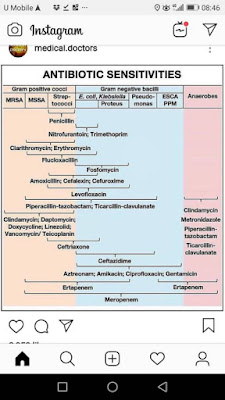
Antibiotics:
- - IV Tazocin 4.5g QID (consist of piperacillin and tazobactem)
- - IV clindamycin 600-900mg TDS
- inhibitor of protein synthesis and protein m synthesis
- effective for gram +ve
- or Cefotaxime + metronidazole
- - unasyn (beta lactam, inhibiting bacterial cell wall synthesis)
- - ciprofloxacin : covers gram negative
Reference:
Monday, November 15, 2021
HPB Q&A for HO
HPB = Hepatobiliary
Anatomy of HPB
Common classifications used in HPB
1. Ranson criteria
2. Mirrizi classification
biliary colic -> acute cholecystitis -> acute cholangitis, pancreatitis, gb empyema and gangrenous gb
presentation of each spectrum
Causes of obstructive jaundice
painless vs painful jaundice
- Painful obstructive jaundice is usually related to gallstones, while painless obstructive jaundice tends to be related to tumours.
Q1: If patient came with features of ascending cholangitis, what Investigation you would like to order
- FBC - to monitor TWC, PLT.
- low platelet: sepsis induces/ severe sepsis that causes coagulopathy
- BUSE - urea, creat monitoring
- hyperbilirubinemia can cause hepatorenal syndrome and renal impairment
- severe sepsis that causes organ failure : kidney failure
- LFT: TB, kiver enzymes and albumin level
- especially ALP and direct bilirubin that can help determin if there are any obstructive jaundice
- ABG: any acidosis or resp failure
- CXR first, as we can use it to rule out perforated viscus
- unless obvious jaundice, so less likely perforated PUD
Q2: why we need ultrasound HBS for ascending cholangitis?
- to rv CBD(common bile duct) size, and any dilatation
- any stone present- if there's obstruction , might need urgent intervention to relieve obstruction
- sometimes stone cant be seen as obscured by bowel gas.
- stone is usually in distal CBD - the narrowest part
- and it is at the back of duodemun
- usg wave cannot go through gas
- To rule out liver abscess
- as the presentation of liver abscess is the same: RHC pain, fever and jaundice
Q3: normal size of CBD?
- 3-5mm
- >8mm considered dilated (usually we use >10mm or 1cm as dilated)
Q4: why do we need to rule out liver abscess urgently?
- if huge liver abscess need to drain urgently.
- gold standard tx: percutaneous drainage and antibiotic
- otherwise it will rupture and cause peritonitis --> severe sepsis --> mortality
Q5. if found out patient have ascending cholangitis secondary to choledocholithiasis, how would you manage?
- resuscitate and stabilize the patient first with ABC
- Airway
- Breathing
- Circulation
- insert branula + blood for Ix and blood culture
- fluid resus
- antibiotics
- analgesia
Q5a. what are the choice of antibiotics use for the pt above?
- HBS common organism are
- function of liver: it uses portal circulation,
- it collects blood from gut - all the guy -> return back to liver
- therefore in the GI tract, mainly organism gram negative and anaerobes
- also called BROAD SPECTRUM of negative organisms
- so we need at least 3rd generation cephalosporin
- eg: cefobid / cefoperazone
- to cover anaerobes: metronidazole
Q6. after analgesics what can we do for patient with ascending cholangitis and in pain?
- if Stable, we can try ERCP to remove the stone . if unable to do so we will put stent to drain the obstruction
- ERCP needs sedation, and prone position. therefore difficult to maintain airway
- If patient is unstable , the option is PTBD
Q7. when to do cholecystectomy?
- once sepsis is resolved
- stone not cleared through ERCP
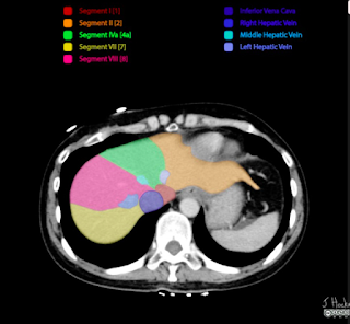
Q8. How do you tell which liver segment was it from a CT scan?
Saturday, November 6, 2021
Electrolyte Imbalance QnA for HO
House Officers must know in ward
- Electrolyte imbalance
Q1: 50year old, 70 kg, male with persistent diarrhea and vomiting for 3days due to food poisoning. He is dehydrated. Vitals are stable, looks lethargic. Na: 128mmol/L
a. How to correct his hyponatremia
- Na deficit (mmol) : (desired Na level - serum Na) x bw x fraction
- 0.6 x kg x (desired Na - serum Na)
- desired Na : 135-145
- Na deficit is 0.6 because extracellular comprises 60%.
- If its potassium (intracellular) therefore 0.4 cause it is 40%
- 0.6 x 70 x (135-128) = 294mmol/L
- infusion = deficit + requirement
- requirement: 1-2mmol/kg/d (we usually use the lower range when calculating)
- infusion: 294 + 70 = 364 mmol/L
- 1 pint NS = 77mmol/ L
- 1L NS = 154mmol/L
- 364 / 77 = 4.7 pints,
- therefore 5 pints of normal saline over 24 hours is needed.
b. the next day his repeated Na is normal: 136,
how do you plan for fluid regime ? for maintenance?
- normally a person need 30-40ml/kg/d (fluid requirement)
- general normal weight for man: 70kg, normal weight for woman : 60kg
- if we take the middle value fluid requirement (35ml/kg/d),
- the man will need 2450ml (around 5 pint as well)
- women is 2100ml, around 4pint.
- 5 pint (2/3 pint NS + 2/3 pint D5%) given to man,
- 4 pint(2 pint NS + 2 pint D5%) given to women.
Q2. what should you watch out during sodium correction?
- Na correction should not exceed 10mmol/L/24 hours
- thats why we use the lower normal when correcting sodium
- even in severe hyponatremia, if Na only 120, your aim of correction is only up to 130, and not up to 135 in 24 hours.
- if there are hypernatremia (rare), mostly we try to find out the associated conditions/ related underlying issues. usually will be given diuretics to excrete excess sodium.
Q3: 50year old, 70 kg, male with persistent diarrhea and vomiting for 3days due to food poisoning. looks lethargic, no ecg changes. K: 3.2mmol/L
a. how to correct his potassium?
- Deficit = (desired K - measured) x Body Weight (BW) x 0.4
- (3.5-3.2) x 70 x 0.4 = 8.4mmol
- 70 for maintanence ,
- maintanence + deficit = infusion
- 70+8.4 = 78.4
- 78.4/13.3 = 5.8gm
- we take normal daily requrement of K : 1mmol/kg/d, therefore we need to convert into gm by dividing 13.3
- 1gm = 13.3mmol
- so we need to correct at least 5 gm first
- can correct with 1g KCL in each pint NS (total 5gm)/24hours
b. what is the maximum/ safe dilution of K+ in 1 pint of NS?
- dilution cannot exceed 40mmol/L
- which also means cannot exceed 20mmol/pint = 20/13.3 = 1.5gm KCl
- we cannot give more than 1.5gm KCl in a pint of NS
c. what is the safe titration of K+?
- titration of potassium cannot exceed 10mmol / hour
- if we need fast correction due to symptomatic,
- max is 1gm KCl in 100cc NS over 1 hour, not faster than that
- If need 2gm infusion, it must be in 2 hours.
- never bolus correction, must always use safe titration
- 2gm KCl infusion in 200cc NS over 2 hours
- OTHERWISE can cause CARDIAC ARREST!
Q4: How do we know the patient has hyperkalemia?
- Cut off point for hyper K:
- K+ > 5.5
- symptomatic ECG changes: tall tented T waves, absence of P wave, broad QRS, PR prolong
- it became dangerous when the ECG changes go towards heart block type of ECG with tall tented T wave --> severe hyperkalemia --> lead to arrythmia then asystole
Q5. what is the fx of each components in lytic coctail?
- lytic cocktail: treatment for hyperkalemia
- Calcium gluconate - 10ml 10% calcium gluconate
- for cardio protection
- Insulin / actrapid 10u :
- drive K+ into cell together with glucose
- Glucose/ D50 - 50ml :
- K+ transporter
- we need to administer calcium gluconate follow by insulin then D50
Q5a. so why we cannot give insulin first instead of glucose?
- Potassium cannot enter the cell by itself, thats why we need glucose first ,since it is the transporter it can allow the insulin to drive the K into the cell.
- GLucose is the transporter
- so we need to load the excess potassium onto the glucose
- Insulin cannot push the K directly, they need transport. therefore they need glucose first, then insulin will push both into the cells.
Q5b. how many times can we give lytic cocktail?
- every 6 hourly
- if the potassium is still not corrected, Hemodialysis (HD) is needed
Q5c. what are the common cause of HyperK in surgery?
- Acute Kidney Injury (AKI)
- severe dehydration, sepsis can cause AKI.
- If there are evidence of sepsis, we need to find the source and remove its source of infection
- antibiotics
- If there are acidosis, we need to find the cause and solve it from there as well.
Q6. what are the common antibiotics used in Gastrointestinal - GI sepsis?
- cefobid and flagyl
- common organism in GI
- gram negative : E.Coli, Klebsiella, Enterobacter
- GI accomodates a broad spectrum of gram negative organisms
- anaerobes
- 1st generation of cephalosporin covers gram positive
- exp: cephalexin
- 2nd generation covers gram positive and a bit of gram negative (narrow spectrum)
- cefuroxime
- 3rd generation covers broad spectrum of gram negative
- cefobid/ cefoperazone
- Metronidazole: covers anaerobes
- If patient has carbuncle, abscess or soft tissue infection / sepsis, the choice of antibiotic should be covering skin organism:
- empirical antibiotic
- penicillin based: cloxacillin, etc.
- for Urology patient, usually have narrow spectrum of organism,
- common: E. Coli
- can use cefuroxime as empirical antibiotic, unless they have sepsis, so may need to consider quinolones to cover for broader/ other pathogens
Q6a. so when do we change antibiotics?
Friday, November 5, 2021
Fluid and Resus common QnA for HO
Part 1: Fluid and its components
Q1. What are the common cause of fluid loss in surgery?
- apparent loss: diarrhea, vomiting and high output stoma
- 3rd space loss:
- loss of water, electrolyte and colloid particles into interstitial space
- which could contribute to edema
- Intestinal obstruction, pancreatitis and ascites
- others: insensible fluid loss (hyperventilation/pyrexia), stress response
Q1a. How patient loss fluids from IO?
- apparent loss: vomiting
- 3rd space loss:
- increased secretions
- bowel obstruction will cause bowel to secrete a lot of secretion to overcome the obstruction.
- mucosal edema, so fluid not absorbed
- so there will be a lot of accumulation of fluid that leads to third space loss
- fluid accumulation in bowel can reach up to 6L
- that could lead to hypovolemic shock
Q1b. How patient loss fluid from pancreatitis?
- systemic inflammation
- inflammation causing release of inflammatory cytokine and other pro-inflammatory mediators,
- leading to capillary leakage
- thus loss of circulatory albumin and fluids to interstitium
- capillary leakage cause fluid shift to third space and then hypotension leads to hypovolemic shock.
Q2. What is the main difference between crystalloid and colloid?
- molecular size
- affects shifting of fluid where low molecular size - low tonicity
Q3. How does fluid moves in human body?
- from low concentration to high concentration
- big molecules fluid (colloids)
- has ability to pull fluids from other compartments : oncotic pressure
- opposite of oncotic pressure: hydrostatic pressure
Q4. What is isotonic?
- a solution concentration that is similar to plasma
- the osmolarity of plasma is around 300 mosm/L
- Normal saline: 308
- Half saline: 154 (hypotonic)
- 3% saline: 1026 (hypertonic)
Q5. What is the difference between Normal Saline (NS) and Hartmann(HM) solution?
- Hartmann has additional potassium, lactate, calcium
- the most "physiological" solution
Q5a. What is the function of lactate in HM
- lactate will be metabolised by liver to HCO3, thus acting as buffer
- especially in met acidosis
Q5b. Can we use HM as resuscitation fluid?
- No
- usually when patient needs resuscitation, already with multiple organ failure or impairment
- so although Hartmann has lactate that could help as buffer, the liver is unable to convert lactate to bicarbonate, which could lead to accumulation
- the accumulation of lactate will worsen the metabolic acidosis
- and Pt in acidosis usually has hyperkalemia, Hartment contains K+ which would further worsen the situation
Q5b. So when do we use hartmann?
- as maintanence, especially in those who need replace electrolyte loss (diarrhea and vomiting)
Q6. What is the function of Dextrose 5%?
- provide hydration
- the have glucose in solution not for calorie to avoid lysis and avoid hypotonic
- calorie in D5: 170/L
- it is just to render solution isotonic once infused in the circulation, once they reach liver will convert into free fluid
- - provide free water that can pass through membrane pores, expanding both intracellular and extracellular spaces
Part 2: Fluid and resuscitations
Q7. 60year old, 70kg man presents with diarrhea and vomiting for 1 week. brought in with hypovolemic shock
Outline your management for this man
- assess airway
- breathing
- circulation
- check the vital signs: unstable/ stable
Q7a : If patient doesn't respond to fluid resus?
- reaccess: if the volume is improved, but patient still hypotensive, he might have other component of shock
- for example: septicaemic shock --> we might need to start inotrope for vasoconstriction
- if cardiogenic shock / has underlying IHD --> get an ECG, and we might need to start with dopamine or dobutamine as the inotropic support.
- if volume is still low,
- can infuse COLLOID to hold the fluid in the circulation
- colloid has oncotic pressure that will hold the fluid intravascularly, thus maintain the BP
- So why we cant give colloid straight away for resus?
- Colloid causes shifting of fluid out of the cell, worsen the hypoperfusion
- in shock, circulation fails and tissue is hypoperfused, if we infuse hypertonic solution all fluid will move from the tissue into the intravascular system .
- Therefore load with volume first (crystalloid)
- resume the circulation
- let them reach the heart, brain and kidney
- after that infuse colloid to hold the volume.
Q8: Define shock
- must have 2 components
- circulatory failure: seen via vital signs
- inadequate tissue perfusion : seen via low SPO2
- sequelae of low perfusion
- multiorgan failure
Q9: Why we cant have central line when patient is in shock?
- it is about the catheter's caliber. the shorter the calibre, faster the infusion.
- if central line, it has long calibre and the rate of infusion is slower
- insertion takes a lot of time
- in shock we need large supply of fluid for the patient
- Poiseuille law
Q10: How do you know patient responded to your fluid resuscitation? what are your AIMS?
Wednesday, October 27, 2021
UGIB Q&A
Q1. What is the anatomical level to ddx upper and lower GI bleed?
- at the duodenal-jejunal junction at the ligament of treitz
- UGIB are bleeding proximal to ligament of trietz
- LGIB can also cause from bleeding of small bowel,
- those bleeding distal to the ligament of treitz can consider as lower GI bleed
Q2. What are your aims in assessment for a patient with UGIB?
- to assess if patient is stable
- if they are in active bleeding
- is it variceal or non variceal bleed
Q2. How to access the stability of the patient?
- vital signs
- mental status
- urine output.
all these could help chart the grade of shock clinically - especially Heart Rate !
- if you notice from the table below.
- the heart rate could already classify the type of shock into class I and class II and it also shows us "THERE is BLEEDING"
- BP came 2nd as when we realise BP drop, shock is already grade 3.
- and blood loss has already reached at least 1.5L
- if grade 2 systolic pressure is still normal and diastolic is low :
Q3. How do we access if they are still actively bleeding?
- colour of hemetemesis
- fresh blood
- coffee ground colour
- amount of hemetemesis
- large?
Q3a. If patient has history of melena, is it usually active bleed or already stopped?
- Yes, usually it is active and has maroon colour, also called fresh melena
- Blood is irritative to gut, they either comes out from the mouth or the anus.
- opposite to fresh melena is old melena or stale melena
- Old melena: black is colour, and towards formed stool.
Q4. Some of the black stool presentation still could be active bleed, what type of presentation we can see in this type of patient?
- large amount of black stool and watery
- also called watery melenic stool
- patient family/ Staff nurse can have complain of patient changing diapers multiple times however soaked. although it is not red, still considered active bleed.
- can happen in both UGIB and LGIB
Rule out UGIB first as 80% of PR bleed is from upper GI, 20% from LGIB
- Despite resuscitation, patient still has persistent tachycardia
Q5. How to access the source of bleeding, variceal vs non variceal?
- Patient's History
- variceal bleed: large amount of fresh hematemesis, has copious blood
- usually described as bowls of blood or cups of blood.
- massive bleeding usually came from varices
- however it is usually PAINLESS
- causes:
- any history of liver disease: jaundice, stigmata of liver disease, portal hypertension secondary to chronic liver disease, hep B
- high risk behaviour
- non variceal bleed: cause is usually peptic ulcer disease (PUD)
- pain
- have hx of epigastric pain
- cause:
- long standing NSAIDS usage/ steroids usage
- hx of hyperuraemia (CKD)
Why do we need to differentiate variceal and non variceal bleed?
- variceal bleed: need urgent OGDS (no active bleed)
- non variceal still can wait and scope within 24 hours
Q1: if patient is suspected with variceal bleed, he has active bleeding and continuously vomit blood.
his vitals are not stable. Outline your acute management
- ABC
- A: If airway compromise to intubate , to secure airway
- insert 2 large bore IV cannula
- blood taking(FBC+coag) + GXM + safe O blood (preferably cross match blood)
- fluid resus with crystalloid then add colloid while waiting for blood and blood products
- can refer to the fluid and electrolyte QnA link here
- to arrest and stop the bleeding
- inserting sengstaken tube
- vital signs monitoring
since patient having active bleed, he is not fit for OGDS, as we couldnt view the source of bleeding clearly. the active bleed will obscure the lens of OGDS
Q1a: what do we do next once patient in ICU- medical therapy?
- start IVI octreotide
- on pt with and without active bleed.
- replace blood loss and correct coagulopathy
- FFP
- start antifibrinolytic agent eg: IV tranxenemic acid 500mg TDS
- transfused if needed
- start PPI infusion
- reduces mortality in variceal bleed
- due to reduce in acid in stomach, it also lower down incidence of rebleeding
- start antibiotic
- as pt usually immunocompromised due to hepatic failure
- in various study shows abx may reduce mortality rate in bleeding pt
- beta blocker
- usually given after patient recover
- as might cause low systemic blood pressure in acute setting
Q2: how does octreotide help with the bleeding?
- it inhibits release of glucagon
- glucagon is splanchnic vasodilator
- when splanchnic circulation is reduced it will cause reduce in portal pressure
Q3: how long can we place the sengstaken tube?
- 48hours
- longest: 72 hours/3days
- as it is just a temporary solution to the problem.
- however have to deflate the esophageal balloon intermittently every 2 hours
- important to reinfate to prevent hypoxia and necrosis of compressed tissue
- and esophageal perforation is fatal cause it is in the thoracic cavity if perforated.
Q4: what is the PPI infusion that is given
- IVI pantoprazole 80mg stat then 8mg/hr
- GOLD STANDARD to give the regime stated above.
- if no pantoprazole, omeprazole is also accepted
Q4b: why PPI is important and superior as medical treatment?
- proton pump inhibitor, blocks the H+/K+ ATPase
- inhibition is irreversible.
- therefore causes profound and prolonged reduction of acid production
CPG malaysia for UGIB mx
Friday, October 1, 2021
China medical universities accredited by malaysia
LIST OF REGISTRABLE QUALIFICATIONS PER SECOND SCHEDULE, MEDICAL ACT 1971
for CHINA universities,
those who are recognised/ listed below can register to practice as houseman in malaysia without taking the examination for provisional registration (EPR)
However those who are not stated above will need to take the exam (examination for provisional registration) before registering to practice housemanship in malaysia.
Reference:
you can cehck the other countries from this link
Thursday, July 8, 2021
QnA sample for OBGYN HO assessment
- when to diagnose gdm ?
- risk factor
- when to check mogtt x1 and x2
- how do you know bsp controlled ? - check hba1c also
- when to induce if GDM d/c and GDM on meds ? ( at how weeks )
- how to start s/c insulin in GDM if needed , and how many unit ?
HTN - Hypertension
- when to diagnose gestational hypertension ?
- when to start antihypertensive in gestational hypertension ?
- when to induce ? ( at how many weeks of gestation)
- which antihypertensive meds to start first ?
- Methyldopa
- Labetolol
- Nifedipine
NVP - Nausea and Vomiting in Pregnancy
- What indicator show that we need to admit this patient if she come with NVP ?
- how to ddx with hyperemesis gravidarum
- If urine ketone positive
- How many pints to hydrate patient with NVP ?
- investigations to take?
Miscarriage
- How to diagnose
- types of miscarriage
- Scan findings in miscarriage
- ET thickness and its relation to status of pregnancy
PPH - Post Partum Haemorrhage
- How to Dx, Mx
- Ddx PPH
- Meds to use if Pitocin already max 80u
- How many time can use Hemabate
- What to do if all med fail
Shoulder dystocia
- What Method, when to do it?
- How to do Mc Robert
- What are the HELPPER
- If all fail what to do